Fungalpedia – Note 129 Sulcosporium
Sulcosporium Phook. & K.D. Hyde
Citation when using this entry: Aumentado et al. in prep – Fungalpedia, plant pathogens. Mycosphere.
Index Fungorum, Facesoffungi, MycoBank, GenBank, Fig *.
Ascomycota, Pezizomycotina, Dothideomycetes, Pleosporomycetidae, Pleosporales, Halotthiaceae
Sulcosporium is recognized as a monotypic genus in Halottiaceae based on the 18S small subunit (SSU) rRNA and 28S large ribosomal subunit RNA (LSU) genes and accommodates S. thailandica (Ariyawansa et al. 2015). Sulcosporium shares similarities with Sulcispora, as noted by Shoemaker & Babcock (1989) and Senanayake et al. (2015). However, Sulcispora belongs to Phaeosphaeriiaceae and is unrelated to Sulcosporium. The ascomata of this genus are solitary, scattered, globose to subglobose, and embedded in the host. The ascomata are uni-loculate and membranous, with a small central papilla that erupts over the host surface, forming an ostiole. The peridium is thin and consists of numerous layers of thick-walled, brown to dark brown, pseudoparenchymatous cells organized in a textura angularis. The hamathecium comprises dense pseudoparaphyses. These pseudoparaphyses anastomoses at the apex and are immersed in a mucilaginous matrix. The asci are 8-spored, bitunicate, and fissitunicate. They are broadly fusiform to clavate, saccate, or ampulliform, with a short blunt pedicel and a rounded apex that forms a completely developed ocular chamber. The ascospores are arranged in overlapping rows of two to three layers (bi- to tri-seriate). Initially, they are hyaline, but as they mature, they become very pale brown. The ascospores are ellipsoidal to fusiform, or clavate, and have a single septum. The asexual morph is undetermined (Ariyawansa et al. 2015).
Sulcosporium causes necrotic leaf spots on Axonopus compresus (Ariyawansa et al. 2015). The leaf spots typically measure 3–5 cm in length and are commonly found on leaf edges. The lesions are irregular shapes and appear pale brown to brown which are discernible due to their distinct reddish-brown boundaries that separate them from the unaffected portion of the leaf (Ariyawansa et al. 2015). It can be likened to Mixtura which incites leaf spots on Poaceae (Eriksson & Yue 1990). Sulcosporium may bear a resemblance to Mixtura in possessing pseudoparaphyses with saccate asci, as mentioned by Eriksson & Yue (1990) and Phookamsak et al. (2014). Nonetheless, Sulcosporium can be distinguished from Mixtura by its two-celled ascospores, whereas Mixtura has muriform ascospores (Ariyawansa et al. 2015) and is a monotypic genus in Phaeosphaeriaceae. Further studies on Sulcosporium are needed considering little information on its etiology and pathogenicity.
Type species: Sulcosporium thailandica Phookamsak & K.D. Hyde
Other accepted species: This genus is monotypic.
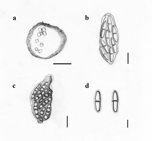
Figure 2. Sulcosporium (redrawn from Ariyawansa et al. 2015). a Vertical section through an ascoma. b, c Asci. d Ascospores. Scale bars: a = 50 μm, b, c = 20 μm, d = 10 μm.
References
Ariyawansa HA, Hyde KD, Jayasiri SC, Buyck B et al. 2015 – Fungal diversity notes 111–252: taxonomic and phylogenetic contributions to fungal taxa. Fungal diversity 75, 27–274.
Eriksson OE, Yue J 1990 – Notes on bambusicolous pyrenomycetes. Mycotaxon 38(1–10), 201–220.
Phookamsak R, Liu JK, McKenzie EHC, Manamgoda DS et al. 2014 – Revision of Phaeosphaeriaceae. Fungal Diversity 68, 159–238.
Senanayake IC, Maharachchikumbura SSN, Hyde KD, Bhat JD et al. 2015 – Towards unraveling relationships in Xylariomycetidae (Sordariomycetes). Fungal Diversity 73(1), 1–85.
Shoemaker RA, Babcock CE. 1989 – Phaeosphaeria. Can J Bot 67, 1500–1599.
Entry by
Herbert Dustin R. Aumentado, Center of Excellence in Fungal Research and School of Science, Mae Fah Luang University, Chiang Rai, Thailand
Edited by Kevin D. Hyde & Ruvishika S. Jayawardena
Published online 5 September 2023