Fungalpedia – Note 286, Rodentomyces
Rodentomyces Doveri, Pecchia, Sarrocco & Vannacci
Citation when using this entry: Perera et al. 2024 (in prep) – Fungalpedia, genera described in 2016.
Index Fungorum, Facesoffungi, MycoBank, GenBank, Fig. 1
Classification: Nectriaceae, Hypocreales, Hypocreomycetidae, Sordariomycetes, Pezizomycotina, Ascomycota, Fungi
Doveri et al. (2010) introduced the monotypic genus Rodentomyces with R. reticulatus as the type, based on the phylogenetic analysis of ITS sequences. Rodentomyces reticulatus occurs in small rodent dung (Doveri et al. 2010). However, the genus name was not valid because the holotype was cited as ‘cultura viva’ in the original protologue (Doveri et al. 2010). Later, the name was validated in the Index Fungorum (2016). The genus is characterized by superficial, astromatic, ostiolate, and pale yellow to luteous ascomata that do not change colour in KOH. The peridium is less than 10 μm thick and is composed of thick-walled cells with angularis texture. Periphyses and apical paraphyses are present. Asci are unitunicate and inamyloid with an inconspicuous apical apparatus. Ascospores are yellowish brown, 1-septate to muriform, tuberculate, and subreticulate with no germ pores or slits (Doveri et al. 2010).
Type species: Rodentomyces reticulatus Doveri, Pecchia, Sarrocco & Vannacci
Other accepted species: This genus is monotypic
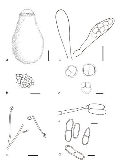
Figure 1 – Rodentomyces reticulatus (DSM 23301, holotype). a Perithecium in water. b Surface view of the peridium cells. c Asci. d Ascospores in different stages of episporial wall deposition. e Conidiophores on water agar. f An empty conidiogenous cell and two mature conidia. g One and two-celled conidia with truncate, eccentric hilum. Scale bars: a, b = 35 μm, c = 25 μm, d = 13 μm, e = 25 μm, f, g = 5 μm. Redrawn from Doveri et al. (2010).
Reference
Entry by
Rekhani Hansika Perera, Center of Excellence in Fungal Research, Mae Fah Luang University, Chiang Rai, 57100, Thailand.
(Edited by Kevin D. Hyde, Samaneh Chaharmiri-Dokhaharani, & Achala R. Rathnayaka)
Published online 28 May 2024