Fungalpedia – Note 285, Pseudosporidesmium
Pseudosporidesmium K.D. Hyde & McKenzie
Citation when using this entry: Perera et al. 2024 (in prep) – Fungalpedia, genera described in 2016.
Index Fungorum, Facesoffungi, MycoBank, GenBank, Fig. 1
Classification: Pseudosporidesmiaceae, Xylariales, Xylariomycetidae, Sordariomycetes, Pezizomycotina, Ascomycota, Fungi
Su et al. (2016) observed that the morphology of Sporidesmium knawiae (Crous et al. 2008) is distinct from other Sporidesmium species. Therefore, the hyphomycetous genus Pseudosporidesmium was established (in Xylariomycetidae, incertae sedis) to accommodate S. knawiae (as Pseudosporidesmium knawiae) (Su et al. 2016). The second species, P. lambertiae, was added, and the genus was placed in the new family Pseudosporidesmiaceae by Crous et al. (2017). Pseudosporidesmium is characterized by black, erumpent, and sporodochial colonies. The mycelium is composed of branched, septate, pale brown, and smooth-walled hyphae. Conidiophores are single or aggregated in groups, erect or somewhat repent, emerging from creeping hyphae or aggregated in black sporodochia. Conidiophores are separated from hyphae by a basal septum; the base is usually not swollen and lacks rhizoids. Conidiophore stipes are cylindrical, brown, smooth, thick-walled, sometimes geniculate, branched in the upper part, or regenerating percurrently. Conidiogenous cells are terminal, cylindrical, brown, and proliferate once percurrently at the apex with apparent flared collarette. The conidiophore grows in length before creating the next conidium. Conidia are solitary, acrogenous, obclavate, 4-euseptate, smooth walled, tapering towards the subobtuse apex, and truncate at the base. Basal cell of the conidia is pale brown, while second to fourth cells are medium brown, and apical cell is pale brown. The hilum has a minute marginal frill. Sometimes, conidia with delayed secession are evident, giving the appearance of lateral conidiogenous loci on conidiogenous cells. The sexual morph is undetermined (Su et al. 2016; Crous et al. 2017). Pseudosporidesmium can be distinguished from Sporidesmium s. str. with molecular support, and conidiophores with percurrent rejuvenation (Su et al. 2016; Crous et al. 2017). Species in this genus are associated with the leaves of Encephalartos lebomboensis and Lambertia formosa (Su et al. 2016; Crous et al. 2017).
Type species: Pseudosporidesmium knawiae (Crous) K.D. Hyde & McKenzie
Other accepted species:
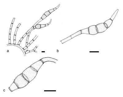
Figure 1 – Pseudosporidesmium knawiae (CBS H-20158, holotype). a Conidiophores with terminal conidiogenous cells. b Conidiogenous cells and conidia. Scale bars: a–c = 10 μm. Redrawn from Crous et al. (2008).
References
Entry by
Rekhani Hansika Perera, Center of Excellence in Fungal Research, Mae Fah Luang University, Chiang Rai, 57100, Thailand.
(Edited by Kevin D. Hyde, Samaneh Chaharmiri-Dokhaharani, & Achala R. Rathnayaka)
Published online 28 May 2024