Fungalpedia – Note 260, Pseudopithomyces
Pseudopithomyces Ariyaw. & K.D. Hyde.
Citation when using this entry: Aumentado et al. 2024 (in prep) – Fungalpedia, plant pathogens.
Index Fungorum, Facesoffungi, MycoBank, GenBank, Fig. 1.
Classification: Didymosphaeriaceae, Pleosporales, Pleosporomycetidae, Dothideomycetes, Pezizomycotina, Ascomycota, Fungi
Pseudopithomyces was established by Ariyawansa et al. (2015) in Didymosphaeriaceae, with Pseudopithomyces chartarum as its type species. This classification system was introduced to encompass various strains previously classified as Pithomyces chartarum (Berk. & M.A. Curtis) M.B. Ellis, P. maydicus (Sacc.) M.B. Ellis, P. sacchari (Speg.) M.B. Ellis and other related Pithomyces strains grouped within the family Didymosphaeriaceae (Ariyawansa et al. 2015). Pseudopithomyces resembles Pithomyces in having echinulate or fusiform, verruculose dark conidia, and the production of brown to black colonies on the host. However, Pithomyces distinguishes itself by producing lighter obovate to oblong verruculose to spinulose conidia, and forming whitish to yellowish colonies on the host (Ariyawansa et al. 2015, Pratibha & Prabhugaonkar 2015). Furthermore, Pseudopithomyces demonstrates a phylogeny distinct from Pithomyces, which displays a polyphyletic distribution across families within the order Pleosporales, such as Didymellaceae and Astrosphaeriellaceae (da Cunha 2014, Ariyawansa et al. 2015, Pratibha & Prabhugaonkar 2015). There are 13 recognised species of Pseudopithomyces based on morphology and molecular data utilising a consolidated set of sequence data sets including ITS, LSU, SSU, gapdh, tef1-α, and rpb2 (Ariyawansa et al. 2015, Crous et al. 2016, Wanasinghe et al. 2018, Jayasiri et al. 2019, Tennakoon et al. 2021). Pseudopithomyces is characterised by micronematous, semi-macronematous, or mononemous conidiophores, and a flexuous, thin-walled, hyaline to subhyaline appearance. Conidiophores are typically unbranched and densely clustered, arise perpendicularly from repent hyphae, and are either monoblastic or blastic. Conidiogenous cells are also closely aggregated, either globose or subglobose, integrated, terminal, determinate, and disintegrate upon maturity to release the conidia. Conidia are acrogenous, dark fuligineous, and vary in septation from single- to multiseptate, either fusiform or subglobose, with verruculose to echinulate surfaces. Conidia are thin-walled, ornamented, and irregularly wide in the middle (Ariyawansa et al. 2015, Crous et al. 2016).
Pseudopithomyces species are associated with diseased or dead leaves, plant stems, and humans, either as saprobes or pathogens. Pseudopithomyces species have been documented on diverse substrates, including on seeds of Medicago sativa (da Cunha 2014), leaves of Acoelorrhaphe wrightii (Ariyawansa et al. 2015), deceased stems of Gnidia polycephala (Crous et al. 2016) and aerial spines of Rosa canina (Wanasinghe et al. 2018), on Entada phaseoloides (Jayasiri et al. 2019), Chromolaena odorata (Mapook et al. 2020), and Morus australis (Tennakoon et al. 2021), Pandanus amaryllifolius (Tibpromma et al. 2018), Triticum aestivum (Perelló et al. 2017). As a plant pathogen, Pseudopithomyces species incite leaf spots on Fragaria × ananassa (Samaradiwakara et al. 2022), Prunus spinosa (Atashi & Fotouhifar 2022), and Tetrapanax papyrifer (Wu et al. 2023), correspondingly proving their pathogenicity. Nevertheless, further studies on Pseudopithomyces are needed, considering the limited information on its etiology and pathogenicity.
Type species: Pseudopithomyces chartarum (Berk. & M.A. Curtis) Jin F. Li, Ariyaw. & K.D. Hyde
Other species: (Species Fungorum – search Pseudopithomyces)
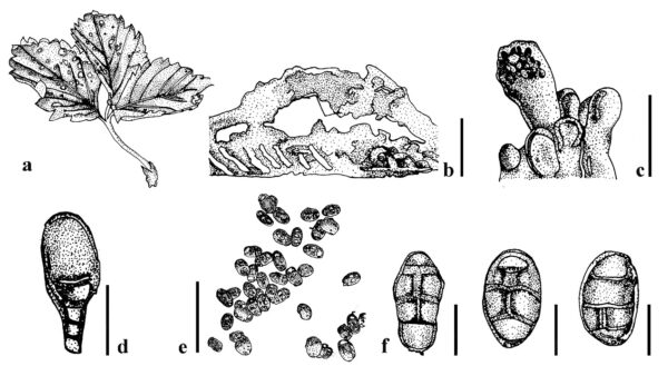
Figure 1 – Pseudopithomyces spp. a Symptoms P. maydicus on leaves of Fragaria × ananassa. b Section of pycnidium. c, d Developing conidia. e P. maydicus conidia. f P. rosae conidia. Scale bars: b = 100 µm; c–f = 10 µm. Redrawn from Wanasinghe et al. (2018) and Samaradiwakara et al. (2022).
References
Entry by
Herbert Dustin R. Aumentado, Center of Excellence in Fungal Research and School of Science, Mae Fah Luang University, Chiang Rai, Thailand
(Edited by Ruvishika S. Jayawardena, Kevin D. Hyde, Samaneh Chaharmiri-Dokhaharani, & Achala R. Rathnayaka)
Published online 21 May 2024