Fungalpedia – Note 148 Fusiformispora
Fusiformispora Phukhams. & K.D. Hyde
Citation when using this entry: Shah et al. (in prep) – Fungalpedia, new genera in 2020. Mycosphere
Classification: Amniculicolaceae, Pleosporales, Pleosporomycetidae, Dothideomycetes, Pezizomycotina, Ascomycota
Index Fungorum, Facesoffungi, MycoBank, GenBank Figs 1, 2.
The monotypic Fusiformispora was introduced by Phukhamsakda et al. (2020). It was isolated as a saprobe from a decaying branch of Clematis fulvicoma found in Chiang Rai Province, Thailand. Its sexual morph is characterized by immersed, scattered or sparsely distributed ascomata. It has a false covering that appears as black or brown, leather-like, globular spots. It contains centrally located ostioles that bear brownish papillae. The peridium contains several layers; the inner layers translucent, cellular of textura angularis. The hamanthecium are closely packed, branched, and filamentous with transverse septa. Fusiformispora possesses bitunicate or fissitunicate asci, having eight spores with thick walls. Asci may be cylindrical, club-like, and rounded apically, with a reduced ocular-type chamber with a bifurcate pedicel. Ascopores are hyaline, biseriately arranged or overlapping, filamentous and tapered at the narrow ends. Each cell may possess one or two small rounded vesicles with a smooth wall, constricted at the septa, and with a fine mucilaginous sheath. The asexual morph is undetermined. Fusiformispora may have similar characteristics to other genera in Amniculicolaceae in having globular, leather-like, brownish ascomata with papillate ostioles and pseudoparaphyses and filamentous, septate, mucilaginous ascospores (Liew et al. 2000, Zhang et al. 2009). Fusiformispora with its terrestrial habitat shows contrasting characteristics from Amniculicola of Amniculicolacea in having barrel or club-shaped asci, and biseriately arranged ascospores. Fusiformispora was identified as a new genus in Amniculicolacea based on the morphology and multiple gene analysis of LSU, SSU, ITS, tef1 and rpb2 sequence data.
Type species: Fusiformispora clematidis Mapook & K.D. Hyde

Figure 1 – Morphology of Fusiformispora clematidis (redrawn from Phukhamsakda et al. 2020). a Section through ascoma. b, c-e, f Ascospores. d Asci. Scale bars: a = 100 μm, b, c-e, f = 20 μm, d = 50 μm
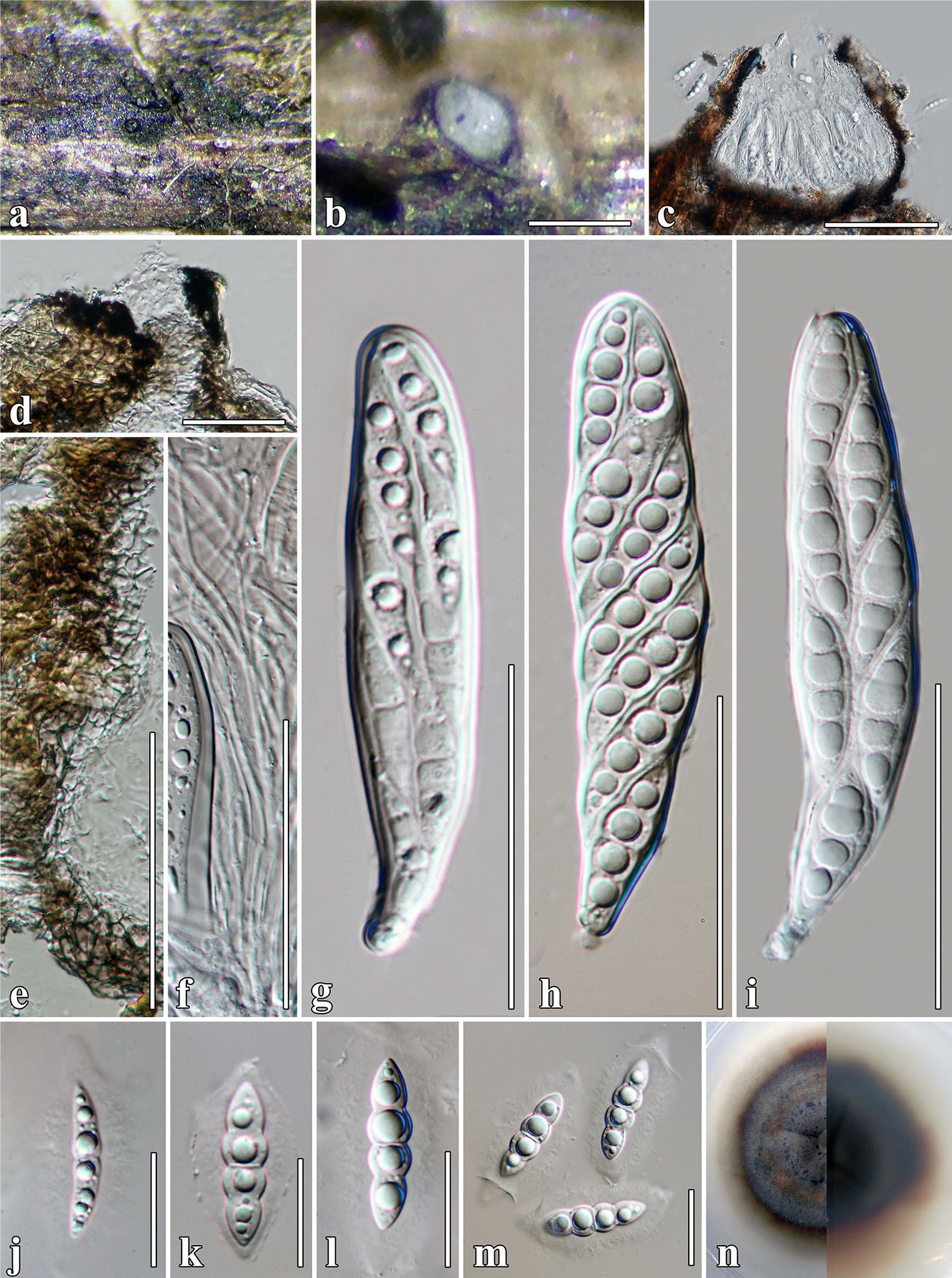
Figure 2 – Fusiformispora clematidis (MFLU 17–1485, holotype). a, b Ascoma on host surface. b c Ascoma section. d Ostiolar canal. e Peridium. f Pseudoparaphyses. g–i Asci. j–m Ascospores. n Culture characteristics. Scale bars: b = 200 μm, c = 100 μm, d, j–m = 20 μm, e–i = 50 μm
References
Entry by
Sujit Shah, Center of Excellence in Fungal Research, Mae Fah Luang University, Chiang Rai 57100, Thailand.
Edited by Roberto Farias & Kevin D. Hyde
Published online 5 October 2023