Fungalpedia – Note 227, Diamantinia
Diamantinia A.N. Mill., Læssøe & Huhndorf.
Citation when using this entry: Doilom et al. 2024 (in prep) – Fungalpedia, Xylariomycetidae.
Index Fungorum, Facesoffungi, MycoBank, GenBank, Fig. 1
Classification: Xylariaceae, Xylariales, Xylariomycetidae, Sordariomycetes, Pezizomycotina, Ascomycota, Fungi
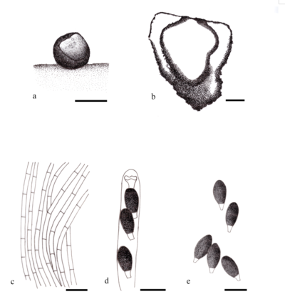
Diamantinia was established based on the morphology by Miller et al. (2003), with D. citrina as the type species which was collected from decaying decorticated wood and corticated branches on the ground in dry deciduous shrubby vegetation in Bahia, Brazil. This monotypic genus is characterized by black, superficial, turbinate stromata, surface minutely roughened, filiform paraphyses, 8-spored, cylindrical asci, amyloid, and apical ring shallow with flaring margins. Ascospores are broadly fusiform, initially hyaline, one-celled, and becoming two-celled by the formation of a transverse septum; upper cell consisting of a swollen, brown head with a narrow, longitudinal germ slit; lower cell consisting of a small, conical, hyaline pedicel (Miller et al. 2003).
Based on the preliminary morphological examination, it can be inferred that D. citrina belongs to Lasiosphaeriaceae due to its filiform paraphyses and apiosporous ascospores, which bear a striking resemblance to those of Strattonia Cif. emend. Lundq. [i.e., Strattonia carbonaria (Phill. & Plowr.) Lundq.]. According to Miller et al. (2003), Diamantinia is placed under Xylariales based on the analysis of partial 28S nuclear ribosomal large subunit DNA. Subsequent morphological examination revealed both an amyloid ring and ascospores with germ slits, which are characteristics of members of the Xylariaceae. On the basis of the keys provided by Barr (1990) and Hawksworth et al. (1995) Diamantinia is classified within the Xylariaceae. However, its paraphyses shape (cylindrical versus tapering), apical ascus ring, ascospores, and peridial anatomy differ to some extent from those of other members of this family (Jensen 1985). Additionally, Diamantinia did not cluster with other Xylariaceae species due to bicellular spores. Miller et al. (2003) suggested a placement of Diamantinia as Xylariales incertae sedis. This taxonomic placement was performed by Lumbsch & Huhndorf (2010), Hyde et al. (2020), and Wijayawardene et al. (2020, 2022).
Type species: Diamantinia citrina A.N. Mill., Læssøe & Huhndorf
Figure 1– Diamantinia citrina. a Vertical section through the stroma and ascomata. b Longitudinal section through stroma showing stromal wall, endostroma (mostly disintegrated in upper part), and ascomal wall. c Paraphyses. d Ascus apices showing amyloid rings in Melzer’s reagent. e Ascospores. Scale bars: a = 500 µm, b = 100 µm, c–e = 10 µm. Redrawn from Miller et al. (2003).
References
Jensen JD. 1985 – Peridial anatomy and pyrenomycete taxonomy. Mycologia 77 (5), 688–701.
Entry by
Mingkwan Doilom, Innovative Institute for Plant Health/ Key Laboratory of Green Prevention and Control on Fruits and Vegetables in South China, Ministry of Agriculture and Rural Affairs, Zhongkai University of Agriculture and Engineering, Guangzhou, 510225, China.
(Edited by Kevin D Hyde, Samaneh Chaharmiri-Dokhaharani, & Achala R. Rathnayaka)
Published online 1 April 2024
