Fungalpedia Note 338, Coprotus
Coprotus Kimbrough and Korf
Citation when using this entry: Thilanga et al. 2024 (in prep) – Fungalpedia, Dung Fungi.
Index Fungorum, Facesoffungi, Mycobank, GenBank, Fig. 1
Classification: Incertae sedis, Pezizales, Pezizomycetidae, Pezizomycetes, Pezizomycotina, Ascomycota, Fungi.
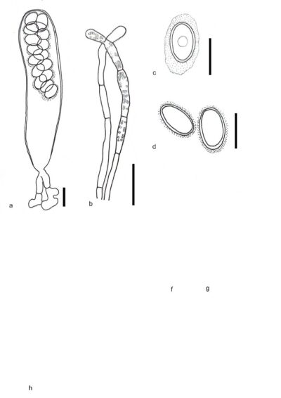
Coprotus was validated by Kimbrough & Korf (1967), encompassing certain species of Ascophanus and Ryparobius Boud. Coprotus was placed in the tribe Theleboleae (family Pezizaceae) by Kimbrough & Korf (1967). The species of this genus are saprobic in the dung of various wild and domestic animals, mainly herbivores. Coprotus is characterized by oblate to lenticular or discoid, glabrous, translucent, or whitish to yellow apothecia with a coprophilous ecology. Anamorph is not known. Asci are functionally operculate, non-amyloid, eight- to 256-spored, producing hyaline, smooth, eguttulate ascospores containing gaseous inclusions referred to as de bary bubbles when placed in anhydrous conditions. Paraphyses are present. They are filiform, mostly bent to uncinate or swollen at the apex, hyaline, or contain pigment. The excipulum is primarily composed of globose to angular cells (Kimbrough et al. 1973). The type species Coprotus sexdecimsporus was sequenced for the first time to ascertain the phylogenetic position of the genus. Which species is difference of other species appeared with disc-lent of apothecial shape, variation of whitish to yellowish pigmentation, elongated marginal cell structure and ellipsoid – narrowly ellipsoid ascospore characters. The phylogenetic analyses performed based on LSU, and concatenate analysis of LSU and ITS, firmly nested the Coprotus species in the Pezizales, as a member of the Boubovia-Coprotus lineage inside the Pyronemataceae (Kušan et al. 2018). There are 31 species listed in Index Fungorum (2024) under Coprotus.
Type specie: Coprotus sexdecimsporus (P. Crouan & H. Crouan) Kimbr. & Korf.
Other species: (Species Fungorum – search Coprotus)
Figure 1 – Coprotus sexdecimsporus a Ascus with ascogenous cells. b Paraphyses. c Freshly ejected ascospore with a sheath. d Mature ascospores. Scale bars: a – d =10 µm. Redrawn from Kušan et al. (2018).
References
Entry by
Dilan Thilanga, Center of Excellence in Fungal Research, Mae Fah Luang University, Chiang Rai, Thailand
(Edited by Chayanard Phukhamsakda,Kevin D. Hyde, Samaneh Chaharmiri-Dokhaharani, & Achala R. Rathnayaka)
Published online 27 August 2024
