Fungalpedia – Note 71 Brunneodinemasporium
Brunneodinemasporium Crous & R.F. Castañeda
Citation when using this data: Huanraluek et al. Fungalpedia, coelomycetes
Index Fungorum, Facesoffungi, MycoBank, GenBank, Coleomycetes.org, Fig1.
Brunneodinemasporium was established by Crous et al. (2012) and included in Chaetosphaeriaceae (Sordariomycetes, Chaetosphaeriales) by phylogenetic analyses based on ITS and LSU gene regions. The genus has a distinctive appearance. Conidiomata are dark brown to black, stromatic, scattered or aggregated, superficial, unilocular and setose and has brown to black, abundant, subulate to cylindrical, simple, septate, unbranched, smooth-walled setae, arising randomly throughout basal stroma. The basal stroma is composed of thick-walled cells of textura angularis. Hyaline to pure brown, cylindrical, branched, thin and smooth-walled conidiophores line the basal stroma in a dense layer. Conidiogenous cells are hyaline, phialidic, subcylindrical to lageniform, determinate, smooth-walled, with a periclinal thickening towards apex when mature. Conidia are hyaline to pale brown, fusiform, with an obtuse apex and truncate base, gently curved or straight, unicellular, bearing single, unbranched, flexuous, tubular appendages at each end. (Crous et al. 2012, Lu et al. 2016, Li et al. 2020, Wu & Diao 2022). The type species of this genus is Brunneodinemasporium brasiliense from a decaying leaf in Brazil (Crous et al. 2012). Another species, B. jonesii was found on decaying wood in a freshwater stream in China (Lu et al. 2016), that is characterized by hyaline to pure brown conidia with mucilaginous balls connecting the conidia in short false chains (Lu et al. 2016, Li et al. 2020). Wu & Diao (2022) introduced Brunneodinemasporium sinense from dead leaves of Cyclobalanopsis glauca in China, which has a character that resembles B. brasiliense, however, B. brasiliense differs in producing smaller conidia than B. sinense. (Wu & Diao 2022)
Types species: Brunneodinemasporium brasiliense Crous & R.F. Castañeda 2012.
Other accepted species:
Brunneodinemasporium jonesii Y.Z. Lu, Jian K. Liu & K.D. Hyde
Brunneodinemasporium sinense W.P. Wu & Y.Z. Diao
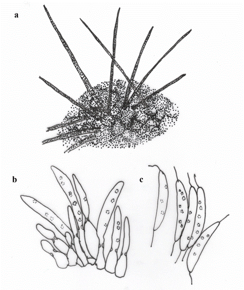
Fig 1 – Brunneodinemasporium brasiliense (redrawn from Crous et al. 2012, Li et al. 2020) a Conidiomatal seta on the surface of conidioma. b Conidiogenous cells and developing conidia. c Conidia. Scale bars: a = 100 µm, b, c = 10 µm.
References
Crous PW, Verkley GJM, Christensen M, Castañeda-Ruiz RF, et al. 2012 – How important are conidial appendages?. Persoonia 28, 126–137.
http://dx.doi.org/10.3767/003158512X652624
Li WJ, McKenZie EHC, Liu JK, Bhat DJ, et al. 2020 – Taxonomy and phylogeny of hyaline-spored coelomycetes. Fungal Diversity 100, 279–801.
https://doi.org/10.1007/s13225-020-00440-y
Lu YZ, Liu KJ, Hyde KD, Bhat DJ, et al. 2016 – Brunneodinemasporium jonesii and Tainosphaeria jonesii spp. nov. (Chaetosphaeriaceae, Chaetosphaeriales) from southern China. 1322–1331.
https://doi.org/10.5943/mycisphere/7/9/6
Wu W, Diao Y. 2022 – Anamorphic chaetosphaeriaceous fungi from China. Fungal Diversity 116, 1–546.
https://doi.org/10.1007/s13225-022-00509-w
Entry by
Naruemon Huanraluek, Center of Excellence in Fungal Research, Mae Fah Luang University, Chiang Rai, Thailand.
(Edited by Kevin D Hyde & Ruvishika S. Jayawardena)
Published online 13 September 2023