Fungalpedia – Note 328, Xenomyrothecium
Xenomyrothecium L. Lombard & Crous
Citation when using this entry: Perera et al. 2024 (in prep) – Fungalpedia, genera described in 2016.
Index Fungorum, Facesoffungi, MycoBank, GenBank, Fig. 1
Classification: Stachybotryaceae, Hypocreales, Hypocreomycetidae, Sordariomycetes, Pezizomycotina, Ascomycota, Fungi
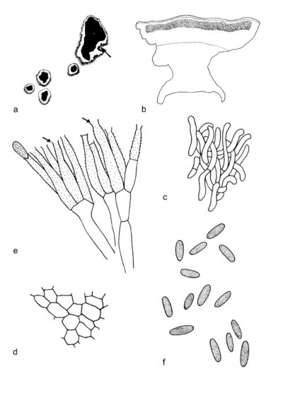
Myrothecium tongaense formed a distinct lineage sister to the Myrothecium s.str. clade and Tangerinosporium thalictricola in the analysis of combined cmdA, ITS, rpb2 and tub2 sequence (Lombard et al. 2016). Lombard et al. (2016) established Xenomyrothecium to accommodate M. tongaense (Xenomyrothecium tongaense). The genus is characterized by stromatic and superficial sporodochial conidiomata. They are irregular in outline, scattered or gregarious and covered by olivaceous green to dark green slimy masses of conidia. The margin is well-developed, slightly involute, intricata in texture, and composed of branched, septate, hyaline, verrucose, and loosely coiled hyphae. The stroma are poorly developed, hyaline and angularis in texture. Conidiophores originating from the stroma had smooth walls, branching, septate, and hyaline. The conidiogenous cells are phialidic, subcylindrical, pale green, verrucose and conspicuously collaretted. Conidia are aseptate, oblong-ellipsoidal, pale green, smooth-walled, with a truncated apex and constricted and truncated base. However, the sexual morph remains undetermined (DiCosmo et al. 1980; Lombard et al. 2016). Xenomyrothecium tongaense occurs on dead thallus of Halimeda (DiCosmo et al. 1980).
Type species: Xenomyrothecium tongaense (W.B. Kendr., DiCosmo & Michaelides) L. Lombard & Crous
Other accepted species: Xenomyrothecium is monotypic.
Figure 1 – Xenomyrothecium tongaense (DAOM 176764, holotype). a Conidiomata on Halimeda sp., arrow indicates fringe of marginal hyphae. b Vertical section of conidioma. c Marginal hyphae. d Cells of the base forming a textura angularis. e Conidiogenous cells, arrows indicate the collarettes. f Conidia. Scale bars: a = ×25, b = ×300, c, d = ×1200, e = ×2000, f = 1200×. Redrawn from DiCosmo et al. (1980)
References
Entry by
Rekhani Hansika Perera, Center of Excellence in Fungal Research, Mae Fah Luang University, Chiang Rai, 57100, Thailand.
(Edited by Kevin D. Hyde, Samaneh Chaharmiri-Dokhaharani, & Achala R. Rathnayaka)
Published online 27 August 2024
