Fungalpedia – Note 308, Varicosporellopsis
Varicosporellopsis Lechat & J. Fourn.
Citation when using this entry: Perera et al. 2024 (in prep) – Fungalpedia, genera described in 2016.
Index Fungorum, Facesoffungi, MycoBank, GenBank, Fig. 1
Classification: Nectriaceae, Hypocreales, Hypocreomycetidae, Sordariomycetes, Pezizomycotina, Ascomycota, Fungi
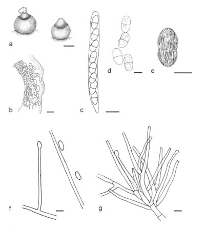
Lechat & Fournier (2016) introduced Varicosporellopsis to accommodate single species, Varicosporellopsis aquatilis, based on LSU and ITS sequence data. Subsequently another two species were added to the genus (Crous et al. 2021; Dou et al. 2023). Varicosporellopsis species were isolated from submerged wood collected in freshwater and sludge in water reservoir (Lechat & Fournier 2016; Crous et al. 2021). The terricolous species Varicosporellopsis shangrilaensis, which was found in a cold, high-altitude, terrestrial environment, occurred in rhizosphere soil (Dou et al. 2023). The sexual morph of the genus is characterized by astromatic, solitary ascomata that are superficial on the substrate with a slightly immersed base. Ascomata are soft-textured, pale orange, and do not change colour in 3% KOH, but becoming very pale yellow to nearly hyaline in lactic acid. They are subglobose and have translucent, broadly conical to rounded apex and a periphysate ostiole. The ascomata collapse laterally upon drying. The surface is covered by tan or hyaline, smooth-walled hyphae. Ascomatal wall comprises two regions. The outer region comprises globose, subglobose to ellipsoidal, pale yellow, thick-walled cells, while inner region comprises hyaline and more flattened cells. The ascomatal surface cells are subglobose and subangular. Asci are unitunicate, cylindrical, short-stipitate, apically truncate to slightly rounded and have a faint apical ring-like thickening. They are associated with moniliform, early deliquescing paraphyses. Varicosporellopsis species produce obliquely uniseriate ascospores that are hyaline to pale yellowish brown and ellipsoid with narrowly to broadly rounded ends. They are 1-septate with a slight constriction at the septum and 2–3 large guttules in each cell. The ascospore wall is roughened with short, sinuous, brown, thick ribs that are sometimes anastomosed (Lechat & Fournier 2016). The asexual morph is hyphomycetous with acremonium-like and/or verticillate conidiophores. Conidiophores are macronematous, mononematous, unbranched or branched at base, 0–2-septate, or reduced to conidiogenous cells, hyaline, flexuous and smooth-walled. The conidiogenous cells are monophialidic with slightly flared collarettes and produce narrowly ellipsoidal to subcylindrical conidia. The verticillate conidiophores emerge laterally from submerged hyphae and produce hyaline to pale salmon coloured, cylindrical, subulate and smooth-walled phialides. The conidia are hyaline, smooth-walled, aseptate, ellipsoidal and attenuated at the base with or without a truncate hilum (Lechat & Fournier 2016; Crous et al. 2021). Varicosporellopsis shangrilaensis produces two types of conidia: reniform macroconidia and spherical microconidia (Dou et al. 2023). Varicosporellopsis differs from Varicosporella in its smaller ascomata and ascospores, and acremonium-like asexual morph (Lechat & Fournier 2016).
Type species: Varicosporellopsis aquatilis Lechat & J. Fourn.
Other accepted species: Species Fungorum – search Varicosporellopsis
Figure 1 – Varicosporellopsis aquatilis (JF15039, holotype). a Ascomata on the substratum. b Lateral ascomatal wall in vertical section. c Ascus. d, e Ascospores. f, g Conidiophores and conidia. Scale bars: a = 100 μm, b, c = 10 μm, d = 5 μm, d, e = 5 μm, f, g = 10 μm. Redrawn from Lechat & Fournier (2016).
References
Entry by
Rekhani Hansika Perera, Center of Excellence in Fungal Research, Mae Fah Luang University, Chiang Rai, 57100, Thailand.
(Edited by Kevin D. Hyde, Samaneh Chaharmiri-Dokhaharani, & Achala R. Rathnayaka)
Published online 8 July 2024
