Fungalpedia – Note 175, Submersispora
Submersispora W. Dong, H. Zhang & K.D. Hyde
Citation when using this entry: Shah et al. 2024 (in prep) – Fungalpedia, new genera in 2020.
Index Fungorum, Facesoffungi, MycoBank, GenBank, Fig 1.
Classification: Longipedicellataceae, Pleosporales, Pleosporomycetidae, Dothideomycetes, Pezizomycotina, Ascomycota, Fungi
This monotypic genus was introduced by Dong et al. (2020) with the type species S. variabilis, which was isolated from decaying wood submerged in a stream in Chiang Mai Province, northern Thailand. Submersispora was introduced as a new genus in Longipedicellataceae, based on morphology and multi-gene phylogenetic analyses (LSU, SSU, ITS, and tef) (Dong et al. 2020). Only the asexul morph is recorded in this genus. Dark, punctiform, and gregarious or scattered or solitary colonies characterize the asexual morph. The mycelium is branched, septate smooth, and thin-walled, hyaline to pale brown, and adheres to the substratum. Conidiophores are semi-macronematous, cylindrical monoonematous, slender, unbranched, septate, smooth, pale brown, and thin-walled. Conidiogenous cells are monoblastic, holoblastic, cuneiform, pale brown, smooth, integrated, and determinate. Conidia are heterogeneous, dispersed, dry, typically subglobose, oblong, or irregular with septa conidia comprise several angular cells ranging in colour from dark black to translucent, depending on maturity (Dong et al. 2020). Both Submersispora and Pseudoxylomyces are freshwater fungal species and share some similar morphological characters, such as holoblastic conidiogenous cells. However, Submersispora species significantly differs from Pseudoxylomyces species by having different-shaped conidia and slender, and semi-macronematous conidiophores (Goh et al. 1997, Phukhamsakda et al. 2016). In this genus, only one species has been listed in Index Fungorum (2023).
Type species: Submersispora variabilis W. Dong, H. Zhang & K.D. Hyde
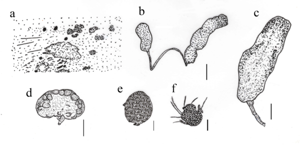
Figure – 1 Morphology of Submersispora variabilis a Colonies on wood surface. b Conidia attached with conidiophores. c Conidiophore, conidiogenous cell, and conidium. d, e Conidia. f Germinated conidium. Scale bars: b-f = 50 μm, c-e = 20 μm. Redrawn from Dong et al. (2020).
References
Entry by
Sujit Shah, Center of Excellence in Fungal Research and School of Science, Mae Fah Luang University, Chiang Rai, 57100, Thailand.
(Edited by Antonio R. Gomes de Farias, Samaneh Chaharmiri-Dokhaharani, & Achala R. Rathnayaka)
Published online 10 January 2024