Fungalpedia – Note 123 Quixadomyces
Quixadomyces Cantillo & Gusmão
Citation when using this entry: Norphanphoun et al., in prep – Fungalpedia, genera and higher taxa of 2022. Mycosphere.
Index Fungorum, Facesoffungi, MycoBank, GenBank, Fig 1.
Based on analysis of combined ITS and LSU sequence data, Crous et al. (2018) introduced Quixadomyces to accommodate Q. cearensis Cantillo & Gusmão, a fungus that was collected from decaying bark in Brazil. However, they did not mention about development of internal structures. The taxonomic placement of Quixadomyces is in Parapyrenochaetaceae (Pleosporales, Dothideomycetes) (Crous et al. 2018). Wanasinghe et al. (2021) introduced Q. hongheensis Wanas. collected from dead twigs of Dodonaea viscosa in China. Quixadomyces hongheensis was introduced based on sequences of concatenated LSU, SSU, ITS, rpb2, ef1α and β-tubulin with strong statistical support (Wanasinghe et al. 2021).
Quixadomyces is characterized by brown to dark brown pycnidia and is known only from its asexual state produced in PDA (Crous et al. 2018, Wanasinghe et al. (2021). The conidiomata are immersed to erumpent, solitary, globose, brown, with a central ostiole, and exuding a hyaline conidial mass. The conidiomata peridium wall has two to three layers of brown cells of textura angularis. Inside the conidiomata are paraphyses, conidiophores, conidiogenous cells, and conidia. Paraphyses are cylindrical, hyaline, and septate. Conidiophores are mostly reduced to conidiogenous cells. Conidiogenous cells are phialidic with periclinal thickening, ampulliform to subcylindrical lining the inner cavity, hyaline, and smooth. Conidia are allantoid with obtuse ends, solitary, hyaline, aseptate and smooth (Fig. 53, Wanasinghe et al. 2021).
Type species: Quixadomyces cearensis Cantillo & Gusmão
Other accepted species:
Quixadomyces hongheensis Wanas.
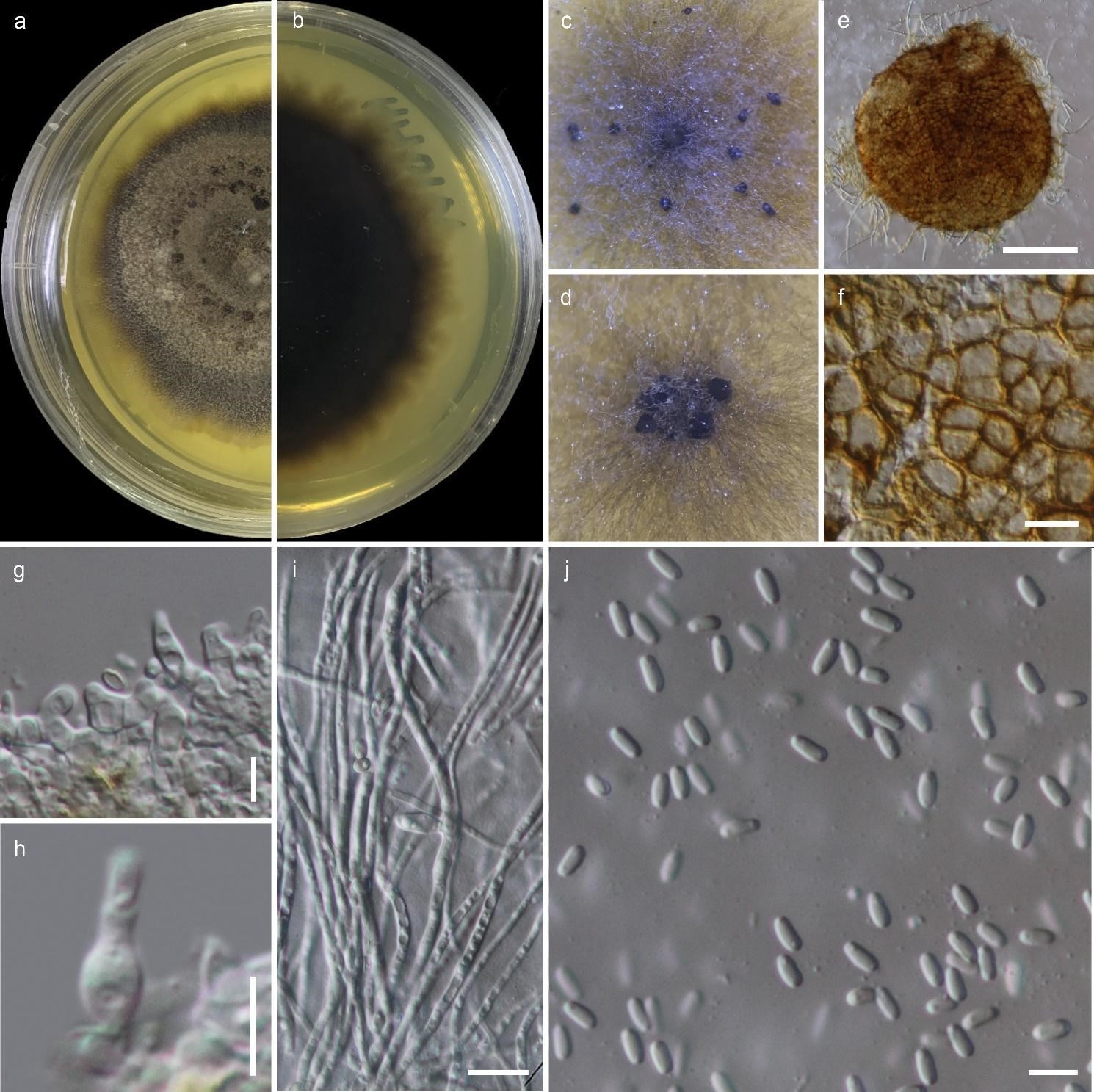
Figure 1 – Quixadomyces hongheensis (KUMCC 20-0215, ex-type culture). a, b Colony on PDA (a from top, b from bottom). c, d Pycnidia on PDA. e Squashed pycnidium. f Pycnidium wall. g, h Conidiogenous cells. i Mycelia. j Conidia. scale bars: e = 200 µm, f, i = 10 µm, g, h, j = 5 µm.
References
Wanasinghe DN, Mortimer PE, Xu J. 2021 – Insight into the systematics of microfungi colonizing dead woody twigs of Dodonaea viscosa in Honghe (China). Journal of Fungi 7, 180.
Entry by
Norphanphoun C., Center of Excellence in Fungal Research, Mae Fah Luang University, Chiang Rai 57100, Thailand and Mushroom Research Foundation, 128 M.3 Ban Pa Deng T. Pa Pae, A. Mae Taeng, Chiang Mai 50150, Thailand
(Edited by Kevin D. Hyde)
Published online 22 September 2023