Fungalpedia – Note 436, Pseudoteratosphaeria
Pseudoteratosphaeria Quaedvl. & Crous
Citation when using this data: Tibpromma et al. 2024 (in prep.) – Fungalpedia, Ascomata.
Index Fungorum, Facesoffungi, MycoBank, GenBank, Fig. 1
Classification: Teratosphaeriaceae, Mycosphaerellales, Dothideomycetidae, Dothideomycetes, Pezizomycotina, Ascomycota, Fungi.
Based on phylogenies of a combined ITS, LSU, RPB2, TEF-1α and TUB alignment, Quaedvlieg et al. (2014) introduced Pseudoteratosphaeria within Teratosphaeriaceae, Mycosphaerellales in Dothideomycetes with the type species Pseudoteratosphaeria perpendicularis. This type of species was initially described as Mycosphaerella perpendicularis by Crous et al. (2006) (≡ Teratosphaeria perpendicularis). While, Pseudoteratosphaeria flexuosa, P. gamsii, P. ohnowa, P. perpendicularis, P. secundaria and P. stramenticola also have been introduced at the same time (Quaedvlieg et al. 2014). Members of this genus can be foliicolous, plant pathogenic or saprobic, morphologically similar to species of Teratosphaeria, and can only be distinguished based on DNA phylogeny. This genus is characterized by having pseudothecial, epiphyllous, black, subepidermal, globose; ostiole central, apical; 2–3 layers of medium brown textura angularis; fasciculate, bitunicate asci, subsessile, obovoid to broadly ellipsoid, slightly incurved, 8-spored; hyaline ascospores with guttulate, thin- walled, fusoid-ellipsoidal, ellipsoidal or obovoid with obtuse ends, medianly 1-septate, widest in the middle of the apical cell, constricted at the septum, tapering towards both ends, but more prominently towards the lower end but no asexual morphs are presently known (Crous et al. 2006, Quaedvlieg et al. 2014).
Type species: Pseudoteratosphaeria perpendicularis (Crous & M.J. Wingf.) Quaedvl. & Crous
Other accepted species: Species Fungorum – search Pseudoteratosphaeria
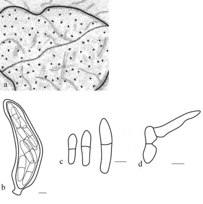
Figure 1 – Pseudoteratosphaeria perpendicularis (CBS H-19691, holotype). a Leaf spot. b Asci. c Ascospores. d Germinating ascospores. Scale bars: b = 3 µm, c-d = 5 µm. Redrawn from Crous et al. (2006).
References
Entry by
Zhang GQ, Center for Yunnan Plateau Biological Resources Protection and Utilization, College of Biological Resource and Food Engineering, Qujing Normal University, Qujing, Yunnan 655011, P.R. China.
(Edited by Saowaluck Tibpromma, Samaneh Chaharmiri-Dokhaharani, & Achala R. Rathnayaka)
Published online 2 December 2024
