Fungalpedia – Note 282, Pseudoanthostomella
Pseudoanthostomella Daranag., Camporesi & K.D. Hyde
Citation when using this entry: Perera et al. 2024 (in prep) – Fungalpedia, genera described in 2016.
Index Fungorum, Facesoffungi, MycoBank, GenBank, Figs. 1 & 2
Classification: Incertae sedis, Xylariales, Xylariomycetidae, Sordariomycetes, Pezizomycotina, Ascomycota, Fungi
Daranagama et al. (2016) established Pseudoanthostomella in Xylariaceae with P. pini-nigrae as the type. The genus was transferred to Xylariales incertae sedis by Samarakoon et al. (2022) based on the combined ITS-LSU-rpb2–tub2–tef1 phylogenies. The sexual morph of Pseudoanthostomella is characterized by immersion in semi-immersed ascomata that developed beneath the clypeus. Ascomata appear as blackened, raised, and conical dome-shaped areas. They are coriaceous or carbonaceous, solitary, rarely aggregated, mostly subglobose with a reduced clypeus, and usually lack a periphysate ostiolar canal. Clypeus is black, thick walled, short, and comprises dark fungal hyphae and host epidermal cells. Peridium with two cell layers; outwardly comprising thick-walled, carbonaceous, compressed, dark brown cells of textura irregularis and inwardly thin-walled, hyaline cells of textura angularis. Paraphyses are less than 5 μm wide at base, shorter than the asci, numerous, filamentous and septate. Asci are 8-spored, unitunicate, broadly cylindrical-clavate with a short-pedicel, apically rounded, and have a discoid-wedged shaped J+ apical ring. The ascospores are arranged in an overlapping, uniseriate manner. They are ellipsoidal, dark brown, smooth walled, and mostly with a conspicuous mucilaginous sheath. Germ slits are either present or rarely absent. If present, it is straight, extending the entire length of the ascospore and positioned on the ventral side. The asexual morph is hyphomycetous with dichotomously branched conidiophores that arise from the hyphae. They are erect, complex, septate, hyaline-light brown and smooth-walled. Monoblastic conidiogenous cells arise in clusters of 3–4, which are discrete, denticulate, and terminal on the branches. Conidia are hyaline, elongated ellipsoidal-fusiform, occasionally curved at the apical end, aseptate, and smooth-walled. Currently, five species are accepted in this genus (Daranagama et al. 2016). Pseudoanthostomella species are saprobic on the dead plant parts of Pinus (Daranagama et al. 2016).
Type species: Pseudoanthostomella pini-nigrae Daranag., Camporesi & K.D. Hyde
Other accepted species: Species Fungorum, search Pseudoanthostomella
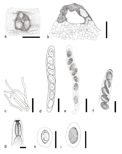
Figure 1 – Pseudoanthostomella pini-nigrae (MFLU 15-3274, isotype),). a Appearance of ascoma on host surface. b Cross section of ascoma. c Paraphyses. d, e Asci. f Ascospores with a wide band. g Apical ring in Melzer’s reagent. h, i Ascospores. Scale bars: a = 200 μm, b = 100 μm, c = 30 μm, d–f, h, i = 20 μm, g = 10 μm. Redrawn from Daranagama et al. (2016)
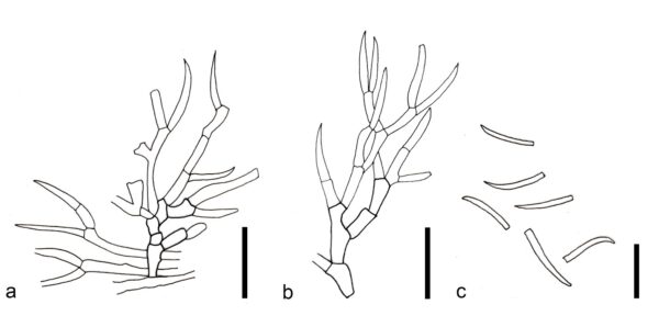
Figure 2 – Asexual morph of Pseudoanthostomella pini-nigrae on OA (MFLUCC 16-0478). a, b Conidiophores with conidiogenous cells. c Conidia. Scale bars: a, b = 20 μm, c = 10 μm. Redrawn from Daranagama et al. (2016)
References
Entry by
Rekhani Hansika Perera, Center of Excellence in Fungal Research, Mae Fah Luang University, Chiang Rai, 57100, Thailand.
(Edited by Kevin D. Hyde, Samaneh Chaharmiri-Dokhaharani, & Achala R. Rathnayaka)
Published online 28 May 2024