Fungalpedia – Note 304, Parengyodontium
Parengyodontium C.C. Tsang, J.F.W. Chan, W.M. Pong, J.H.K. Chen, A.H.Y. Ngan, M. Cheung, C.K.C. Lai, D.N.C. Tsang, S.K.P. Lau & P.C.Y. Woo
Citation when using this entry: Perera et al. 2024 (in prep) – Fungalpedia, genera described in 2016.
Index Fungorum, Facesoffungi, MycoBank, GenBank, Fig. 1
Classification: Cordycipitaceae, Hypocreales, Hypocreomycetidae, Sordariomycetes, Pezizomycotina, Ascomycota, Fungi
Tsang et al. (2016) proposed Parengyodontium for Engyodontium album that formed a distinct phylogenetic lineage within Cordycipitaceae in the analysis of ITS-LSU sequences. Furthermore, E. album chemotaxonomically distinct from other species of Engyodontium (Tsang et al. 2016). Parengyodontium is characterized by cobweb-like, white colonies that are lanose to floccose, velvety to downy and do not diffuse pigment. Hyphae are septate. Conidiophores are upright or procumbent and giving rise to several whorls of conidiogenous cells. Conidiogenous cells have elongated and tapered basal part of and zigzag shaped terminal fertile region. Conidia are hyaline, globose to subglobose, apiculate and smooth-walled. They formed at each bent point of the zigzag rachides. The sexual morph is undetermined (Tsang et al. 2016). Parengyodontium album was isolated from various substrates such as book cover, Pisum sativum, dismal swamp soil and human body (Tsang et al. 2016). The genus currently includes three species (Tsang et al. 2016; Parker et al. 2020; Teixeira et al. 2020).
Type species: Parengyodontium album (Limber) C.C. Tsang, J.F.W. Chan, W.M. Pong, J.H.K. Chen, A.H.Y. Ngan, Cheung, C.K.C. Lai, D.N.C. Tsang, S.K.P. Lau, P.C.Y. Woo
Other accepted species: Species Fungorum – search Parengyodontium.
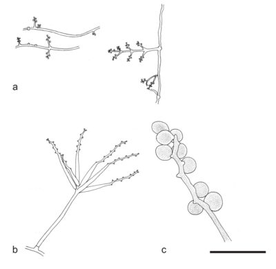
Figure 1 – Parengyodontium album (HKU48). a Conidiogenous cells arranged in whorls of smooth-walled conidiophores. b Whorl of conidiogenous cells (mounted with 5% KOH and calcofluor white stain). c Conidia smooth, hyaline, oval, apiculate, and produced at each bent point of the zigzag rachides. Scale bars: c = 5 μm. Redrawn from Tsang et al. (2016)
References
Entry by
Rekhani Hansika Perera, Center of Excellence in Fungal Research, Mae Fah Luang University, Chiang Rai, 57100, Thailand.
(Edited by Kevin D. Hyde, Samaneh Chaharmiri-Dokhaharani, & Achala R. Rathnayaka)
Published online 8 July 2024