Fungalpedia – Note 320, Paramyrothecium
Paramyrothecium L. Lombard & Crous
Citation when using this entry: Perera et al. 2024 (in prep) – Fungalpedia, genera described in 2016.
Index Fungorum, Facesoffungi, MycoBank, GenBank, Fig. 1
Classification: Stachybotryaceae, Hypocreales, Hypocreomycetidae, Sordariomycetes, Pezizomycotina, Ascomycota, Fungi.
Lombard et al. (2016) introduced Paramyrothecium for a group of myrothecium-like taxa produce 1–3-septate and thin-walled setae that encircle the sporodochia. In their phylogenetic inference based on cmdA, ITS, rpb2 and tub2 loci, Paramyrothecium species showed a distant relationship to Myrothecium s.str. (Lombard et al. 2016). The Paramyrothecium is characterized by scattered or gregarious sporodochial conidiomata, with or without a white setose fringe surrounding an olivaceous green to dark green slimy mass of conidia. Sporodochia are stromatic, superficial, cupulate, oval, or irregular in shape. The stroma of Paramyrothecium is poorly developed or well-developed, hyaline to subhyaline, and consists of hyphae arranged in a globulosa and/or angularis texture. Setae are hyaline, tapered to an acute apex, thin-walled, 1–3(–4)-septate, straight to flexuous, and sometimes sinuous above the apical septum. Macronematous conidiophores are penicillately branched, hyaline, and smooth walled. Conidiogenous cells are phialidic or percurrent, hyaline, sometimes darker at the apex, smooth to lightly verrucose, cylindrical to subcylindrical, and narrower towards the tip. They had conspicuous collarette and periclinal thickening. Conidia are aseptate to 1-septate, cylindrical to ellipsoidal to obovoid, straight to curved and hyaline to pale green with smooth walls. The sexual morph remains undetermined (Lombard et al. 2016). Paramyrothecium species have been reported as plant pathogens that cause leaf spots and leaf blight in diverse plant hosts (Lombard et al. 2016; Armand et al. 2023). Paramyrothecium includes 22 species (Index Fungorum 2024).
Type species: Paramyrothecium roridum (Tode) L. Lombard & Crous
Other accepted species: Species Fungorum, search Paramyrothecium
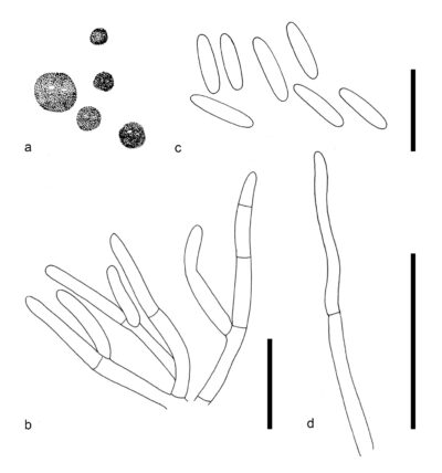
Figure 1 – Paramyrothecium roridum (CBS 357.89, ex-epitype). a Sporodochial conidiomata on SNA. b Seta. c Conidiogenous cells. d Conidia. Scale bars: c–d = 10 μm. Redrawn from Lombard et al. (2016)
References
Current Research in Environmental & Applied Mycology (Journal of Fungal Biology) 13(1), 57–67.
Entry by
Rekhani Hansika Perera, Center of Excellence in Fungal Research, Mae Fah Luang University, Chiang Rai, 57100, Thailand.
(Edited by Kevin D. Hyde, Samaneh Chaharmiri-Dokhaharani, & Achala R. Rathnayaka)
Published online 27 August 2024