Fungalpedia – Note 281, Parafuscosporella
Parafuscosporella Jing Yang & K.D. Hyde
Citation when using this entry: Perera et al. 2024 (in prep) – Fungalpedia, genera described in 2016.
Index Fungorum, Facesoffungi, MycoBank, GenBank, Fig. 1
Classification: Fuscosporellaceae, Fuscosporellales, Hypocreomycetidae, Sordariomycetes, Pezizomycotina, Ascomycota, Fungi
Yang et al. (2016) introduced the hyphomycetous genus Parafuscosporella, with P. moniliformis as the type. The genus was accepted within Fuscosporellaceae based on LSU-SSU-rpb2 phylogenetic analysis (Yang et al. 2016). Parafuscosporella is characterized by effuse, scattered, sporodochial, erumpent, spherical, velvety, and black colonies on natural substrates. The mycelium is partly immersed and partly superficial and composed of septate, hyaline, and pale brown hyphae. Semi-macronematous and mononematous conidiophores arranged compactly. They are flexuous, simple or branched, hyaline, and smooth walled. Conidiophores are mostly moniliform with ellipsoidal, clavate, or globose to subglobose cells. Monoblastic conidiogenous cells give rise to acrogenous conidia. Conidiogenous cells are terminal, integrated, occasionally discrete, globose, subglobose, ellipsoidal or clavate, hyaline, and smooth walled. Parafuscosporella produces ellipsoidal to broadly obpyriform smooth-walled conidia with a septum near the base, and sometimes with a small protuberance. Conidia are dark brown to black, with pale brown basal cells (Yang et al. 2016). The sexual morph is undetermined. Parafuscosporella species are saprobic on twigs in freshwater habitats (Yang et al. 2016, 2020; Boonyuen et al. 2021; Wang et al. 2023).
Type species: Parafuscosporella moniliformis Jing Yang, Bhat & K.D. Hyde
Other accepted species: Species Fungorum – search Parafuscosporella
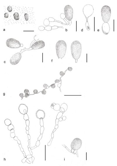
Figure 1 – Parafuscosporella moniliformis (MFLU 15-1161, holotype). a Sporodochia on submerged wood. b, c Conidiophores and conidia. d–f Conidia. g Conidia on PDA. h Conidia and conidiogenous cells on PDA. i Conidium on PDA. Scale bars: a = 500 μm, b–e = 20 μm, f = 15 μm, g = 30 μm, h = 10 μm, i = 5 μm. Redrawn from Yang et al. (2016).
References
Entry by
Rekhani Hansika Perera, Center of Excellence in Fungal Research, Mae Fah Luang University, Chiang Rai, 57100, Thailand.
(Edited by Kevin D. Hyde, Samaneh Chaharmiri-Dokhaharani, & Achala R. Rathnayaka)
Published online 28 May 2024