Fungalpedia – Note 258, Neocordana
Neocordana M. Hern.-Rest. & Crous.
Citation when using this entry: Aumentado et al. 2024 (in prep) – Fungalpedia, plant pathogens.
Index Fungorum, Facesoffungi, MycoBank, GenBank, Fig. 1.
Classification: Pyriculariaceae, Magnaporthales, Sordariomycetidae, Sordariomycetes, Pezizomycotina, Ascomycota, Fungi
Neocordana was introduced by Hernández-Restrepo et al. (2015) to encompass Neocordana musae as the type species. Neocordana has grey to brown colony and has both superficial and submerged mycelium, with smooth, segmented, transparent hyphae that turn brown as they age. The conidiophores are erect or slightly curved, initially dark brown at the base and lighter at the top. The conidiophores are smooth and have a swollen base under the host plant epidermis. The conidiogenous cells are whole and branch out in a tooth-like manner, cylindrical or slightly clubbed, found at both ends and intercalary, smooth, and varying from light brown to brown. The conidia are dry, pale brown, solitary 1-septate, broadly elliptical, elongated, obovoid, club-shaped or pear-shaped, smooth, thick-walled, and have a visibly darker, thickened hilum and appressoria (Hernández-Restrepo et al. 2015).
Neocordana is a phytopathogenic species resembling Cordana. However, these species are more associated with Pyriculariaceae (Magnaporthales) than with Cordanaceae (Cordanales). Pyriculariaceae was defined by Klaubauf et al. (2014), comprising significant plant disease-causing genera, such as Deightoniella (Hughes 1952), and various genera resembling Pyricularia. Deightoniella is differentiated from Neocordana by its reduced conidiophores, appearing as conidiogenous cells with a flattened scar, and its conidia possess a pore at the base (Hughes 1952, Klaubauf et al. 2014), whereas Neocordana displays septate conidiophores featuring tooth-like conidiogenous cells and conidia with a prominent hilum. However, similarities arise between pyricularia-like fungi and Neocordana, notably in brown septate conidiophores with multi-branched tooth-like conidiogenous cells. However, pyricularia-like fungi differ from pyriform to obclavate and 2-septate conidia and are typically isolated from grasses (Seifert et al. 2011, Klaubauf et al. 2014). Conversely, Neocordana has largely ellipsoid, obovoid to pyriform, and 1-septate conidia and demonstrates pathogenicity towards Musa or Canna species (Hernández-Restrepo et al. 2015).
Species Fungorum (2024) are listed seven recognised Neocordana species; N. johnstonii, N. malayensis, N. musae, N. musarum, N. musicola, N. musigena, and N. versicolor based on combined morpho-molecular analysis utilising ITS and LSU gene regions. (Hernández-Restrepo et al. 2015, Crous et al. 2016, 2019, Samarakoon et al. 2019).
Neocordana is a necrotrophic pathogen that only occurs in Musa spp. (Musaceae) and Canna denudata (Cannaceae) leaves (Jones 1999, Hernández-Restrepo et al. 2015, Crous et al., 2016, 2019) and no documented incidence on fruits, roots, or stems. Neither cross-infectivity nor alterations in host behavior have been observed thus far. Leaf spot infections of Musa spp. are prevalent in fields and pose a significant threat to crop yield (Jones 1999, Hernández-Restrepo et al. 2015, Samarakoon et al. 2019). The invasion and penetration of Neocordana pathogens predominantly occurs under humid conditions (Jones 1999). Notably, Neocordana-induced leaf spots on Musa sp. exhibit discernible concentric rings at the centers within necrotic regions (Hernández-Restrepo et al. 2015, Crous et al., 2016, 2019). The lesions appear large, pale brown, oval to fusiform in shape (Jones 1999). Encircling the brown necrotic zones is a distinct bright yellow chlorotic lesion dividing the diseased leaf area from the unaffected portion (Jones 1999). These lesions may manifest similarly in areas previously affected by other pathogens (Samarakoon et al. 2019). The disease symptoms in Canna sp. bear some resemblance to those in Musa spp. (Soares et al. 2005). Symptoms manifest as lesions resembling eyes on the leaf, dispersed extensively across the surface, and ultimately leading to full necrosis of the affected leaf (Soares et al. 2005, Samarakoon et al. 2019). Pathogenicity studies of Neocordana spp. were performed in various studies (Yanping et al. 2021, Sandid et al. 2023) resulting in symptoms as depicted above.
Type species: Neocordana musae (Zimm.) M. Hern.-Rest. & Crous
Other species:
- Neocordana johnstonii (M.B. Ellis) Hern.-Restr. & Crous
- Neocordana malayensis Crous
- Neocordana musarum Crous
- Neocordana musicola Hern.-Restr. & Crous
- Neocordana musigena Crous
- Neocordana versicolor (D.J. Soares & R.W. Barreto) Hern.-Restr. & Crous
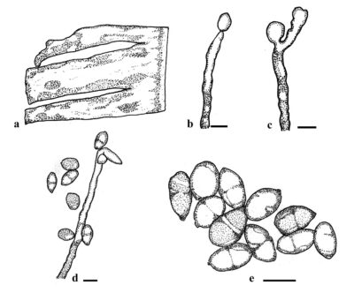
Figure 1 – Neocordana musarum. a Leaf symptoms in Musa sp. leaf. b, c, d Conidiophores with conidia. e Conidia. Scale bars = 10 µm. Redrawn from Crous et al. (2016).
References
Hughes SJ 1952 – Fungi from the Gold Coast. I. Mycological Papers 48, 1–91.
Jones DR. 1999 – Diseases of banana, Abacá, and Enset. CABI Pub, Wallingford, Oxon, UK, New York.
Species Fungorum 2023. Accessed on November 20, 2023, at URL: https://speciesfungorum.org/.
Entry by
Herbert Dustin R. Aumentado, Center of Excellence in Fungal Research and School of Science, Mae Fah Luang University, Chiang Rai, Thailand
(Edited by Ruvishika S. Jayawardena, Kevin D. Hyde, Samaneh Chaharmiri-Dokhaharani, & Achala R. Rathnayaka)
Published online 21 May 2024