Fungalpedia – Note 279, Neoanthostomella
Neoanthostomella D.Q. Dai & K.D. Hyde
Citation when using this entry: Perera et al. 2024 (in prep) – Fungalpedia, genera described in 2016.
Index Fungorum, Facesoffungi, MycoBank, GenBank, Fig. 1
Classification: Incertae sedis, Xylariales, Xylariomycetidae, Sordariomycetes, Pezizomycotina, Ascomycota, Fungi
Dai et al. (2017) introduced Neoanthostomella with its type N. pseudostromatica. Subsequently there are four species added to the genus (Daranagama et al. 2016; Tennakoon et al. 2021). However, after examine the holotype material and based on phylogenetic analysis Samarakoon et al. (2022) confirmed that N. fici and N. viticola clustered within Anthostomella helicofissa strains. Therefore, only two species (N. bambusicola and N. pseudostromatica) can be recognized within Neoanthostomella. The genus is characterized by solitary to gregarious pseudostromata that are circular to elliptical and raised as blacked areas on the host tissue. Two to five ascomata grow together in a single pseudostroma. They are immersed, globose to subglobose, dark brown, coriaceous, with a central, periphysate, ostiolate neck. The peridium is composed of several layers of compressed, brown to hyaline cells of the textura angularis. Hamathecium of dense, long, septate, paraphyses intermixed with asci. The asci are cylindrical with a short furcate pedicel, unitunicate, 8-spored, and lack an apical ring. Ascospores are aseptate, dark brown, guttulate, smooth-walled, uni-to bi-seriate or sometimes overlapping, ellipsoid, and somewhat pointed at the ends. They also have mucilaginous sheaths. The germ slit of the ascospore is straight and extending over the full-length (Dai et al. 2017). The asexual morph is undetermined. Neoanthostomella species are saprobic in dead bamboo plant parts (Dai et al. 2017; Samarakoon et al. 2022).
Type species: Neoanthostomella pseudostromatica D.Q. Dai & K.D. Hyde
Other accepted species:
- Neoanthostomella bambusicola Samarak. & K.D. Hyde.
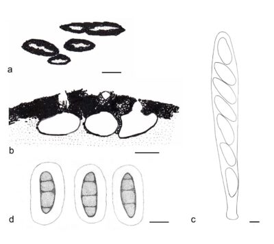
Figure 1 – Neoanthostomella pseudostromatica (MFLU 15–1190, holotype). a Appearance of pseudostromata on bamboo host. b Vertical section of pseudostroma. c Ascus. d Ascospores. Scale bars: a = 1 mm, b = 100 μm, c, d = 5 μm. Redrawn from Dai et al. (2017).
References
Entry by
Rekhani Hansika Perera, Center of Excellence in Fungal Research, Mae Fah Luang University, Chiang Rai, 57100, Thailand.
(Edited by Kevin D. Hyde, Samaneh Chaharmiri-Dokhaharani, & Achala R. Rathnayaka)
Published online 28 May 2024