Fungalpedia – Note 318, Myxospora
Myxospora L. Lombard & Crous
Citation when using this entry: Perera et al. 2024 (in prep) – Fungalpedia, genera described in 2016.
Index Fungorum, Facesoffungi, MycoBank, GenBank, Fig. 1
Classification: Stachybotryaceae, Hypocreales, Hypocreomycetidae, Sordariomycetes, Pezizomycotina, Ascomycota, Fungi.
Lombard et al. (2016) introduced Myxospora to accommodate Myrothecium masonii and a group of myrothecium-like taxa that produce mostly fusiform conidia with an apical hilum and lack funnel-shaped mucoid appendages. In the phylogenetic analysis of cmdA, ITS, rpb2, and tub2 sequences by Lombard et al. (2016), these taxa clustered distant to Myrothecium s. str. clade. Myxospora is characterized by synnematous or sporodochial conidiomata. Synnemata consist of bundles of parallel, longitudinal, and closely compacted hyphae that terminate in whorls of 2–4 conidiogenous cells. Marginal hyphae of the synnemata terminate in hyaline, bulbous, and verrucose cells. Synnemata occur on poorly developed stroma, are cylindrical to pyriform, unbranched, broadened at the apex, and are covered by an olivaceous green to black slimy mass of conidia. Myxospora species produce sporodochia that are stromatic, superficial, scattered or rarely gregarious, oval or irregular in outline, without a white setose fringe surrounding an olivaceous green to dark green slimy mass of conidia. Stromas are poorly developed and hyaline, with an angularis texture. Setae are rare, septate, flexuous, subhyaline, thick-walled, tapered to an obtuse apex that protrudes through the conidial masses. Conidiophores are macronematous, verticillately or penicillately branched, and hyaline with smooth to lightly verrucose walls. Conidiogenous cells are phialidic, hyaline, subcylindrical, and narrow at the tip. They have smooth walls, conspicuous collarettes, and periclinal thickenings. Conidia have smooth walls, are aseptate, fusiform, and hyaline, and sometimes become pigmented with age. The apical hilum of the conidia lack funnel-shaped mucoid apical appendage. The sexual morph remains undetermined (Lombard et al. 2016). Myxospora species are saprophytes or pathogens that can survive in diverse hosts (Lombard et al. 2016; Chen et al. 2023). Myxospora currently accommodates seven species (Lombard et al. 2016; Chen et al. 2023).
Type species: Myxospora masonii (M.C. Tulloch) L. Lombard & Crous
Other accepted species: Species Fungorum, search Myxospora
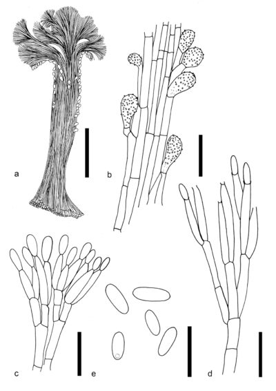
Figure 1 – Myxospora masonii (IMI 158346, holotype). a Synnema. b Marginal hyphae. c, d Conidiophores and conidiogenous cells. e Conidia. Scale bars: a = 100 μm, b–d = 10 μm. Redrawn from Tulloch (1972).
References
Tulloch M. 1972 – The genus Myrothecium. Mycological Papers 130, 1–42.
Entry by
Rekhani Hansika Perera, Center of Excellence in Fungal Research, Mae Fah Luang University, Chiang Rai, 57100, Thailand.
(Edited by Kevin D. Hyde, Samaneh Chaharmiri-Dokhaharani, & Achala R. Rathnayaka)
Published online 27 August 2024