Fungalpedia – Note 473, Hyalocladosporiella
Hyalocladosporiella Crous & Alfenas
Citation when using this data: Tibpromma et al. 2024 (in prep.) – Fungalpedia, Ascomata.
Index Fungorum, Facesoffungi, MycoBank, GenBank, Fig. 1
Classification: Incertae sedis, Chaetothyriales, Chaetothyriomycetidae, Eurotiomycetes, Pezizomycotina, Ascomycota, Fungi
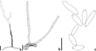
Crous et al. (2014) introduced Hyalocladosporiella with the type species Hyalocladosporiella tectonae and other species H. cannae was introduced by Crous et al. (2017), based on morphological and molecular studies. Hyalocladosporiella is characterized by its dimorphic conidiophores; microconidiophores are subcylindrical, erect, brown, straight to geniculate-sinuous, septate, and macroconidiophores are cylindrical, flexuous, erect, brown, smooth, unbranched, lacking rhizoids, and septate; conidiogenous cells are integrated, terminal, subcylindrical, smooth, brown; loci sympodially arranged, subdenticulate, slightly thickened, and darkened. Primary ramoconidia are fusoid-ellipsoidal to subcylindrical, hyaline to pale olivaceous, guttulate, septate, smooth-walled with thickened and darkened hila, whereas secondary ramoconidia are branched chains, fusoid-ellipsoidal, hyaline, guttulate, with one to three apical loci that are thickened and darkened; intermediary conidia are fusoid-ellipsoid, hyaline, guttulate, and terminal conidia are fusoid-ellipsoid, hyaline, guttulate, loci thickened and darkened (Crous et al. 2014). According to phylogenetic analyses by Colmán et al. (2021), two species of Hyalocladosporiella (Herpotrichiellaceae: Chaetothyriales) are congeneric with Digitopodium and morphologically similar to Digitopodium hemileiae. Therefore, species of Hyalocladosporiella are re-allocated to Digitopodium.
Type species: Hyalocladosporiella tectonae Crous & Alfenas
Current name: Digitopodium tectonae (Crous & Alfenas) A.A. Colmán & R.W. Barreto
Other accepted species: Species Fungorum – search Hyalocladosporiella
Figure 1 – Digitopodium tectonae. a, b Conidiophores and conidia. c Conidia. Scale bars: a-c = 10 μm. Redrawn from Crous et al. (2014).
References
Entry by
Priyashantha AKH, Department of Biology, Faculty of Science, Chiang Mai University, Chiang Mai 50200, Thailand
(Edited by Saowaluck Tibpromma, Samaneh Chaharmiri-Dokhaharani, & Achala R. Rathnayaka)
Published online 3 December 2024
