Fungalpedia – Note 420, Franwilsia
Franwilsia S.Y. Kondr., Kärnefelt, Elix, A. Thell & Hur
Citation when using this data: Tibpromma et al. 2024 (in prep.) – Fungalpedia, Ascomycota.
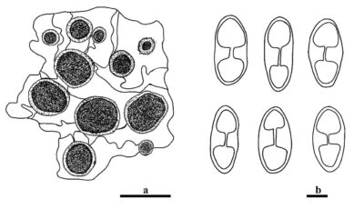
Index Fungorum, Facesoffungi, MycoBank, GenBank, Fig. 1
Classification: Teloschistaceae, Teloschistales, Lecanoromycetidae, Lecanoromycetes, Pezizomycotina, Ascomycota, Fungi.
Franwilsia was introduced by Kondratyuk et al. (2014) and typified with Franwilsia bastowii based on morphology and single gene phylogeny analysis of ITS, LSU, and SSU. Currently, four Franwilsia species (F. bastowii, F. kilcundaensis, F. renatae and F. skottsbergii) are accepted in this genus. They have been isolated from trees and lichens in Australian, Chile, South African and Victoria (Kondratyuk et al. 2009, 2014, 2018). Franwilsia is mainly characterized by whitish to gray or dark gray, cortical layer palisade plectenchymatous, continuous to areolate thallus, zeorine or lecanorine apothecia, subhymenium, lower portion of hymenium, basal portion of true exciple richly by oil droplets or agglomerations, 8-spored asci, polarilocular ascospores, and bacilliform to narrowly bacilliform conidia (Kondratyuk et al. 2014). Morphologically, Franwilsia is similar to Mikhtomia in having a hymenium, subhymenium, and basal portion of true exciples rich in oil droplets (Kondratyuk et al. 2014). However, it differs from the latter in having a leptodermatous paraplectenchymatous true exciple and much larger irregular oil agglomerations in the subhymenium (Kondratyuk et al. 2014). Franwilsia can be distinguished from other genera based on its morphology and phylogeny.
Type species: Franwilsia bastowii (S.Y. Kondr. & Kärnefelt) S.Y. Kondr., Kärnefelt, A. Thell, Elix, J. Kim, A.S. Kondratiuk & Hur
Other accepted species: Species Fungorum – search Franwilsia
Figure 1 – Morphological features of Franwilsia bastowii. a Apothecia. b Ascospores. Scale bars: a = 1 mm, b = 5 µm. Redrawn from Kondratyuk et al. (2009).
References
Entry by
Liu XF, Center for Yunnan Plateau Biological Resources Protection and Utilization, College of Biological Resource and Food Engineering, Qujing Normal University, Qujing, Yunnan 655011, China; Center of Excellence in Fungal Research, Mae Fah Luang University, Chiang Rai 57100, Thailand; School of Science, Mae Fah Luang University, Chiang Rai 57100, Thailand.
(Edited by Saowaluck Tibpromma, Samaneh Chaharmiri-Dokhaharani, & Achala R. Rathnayaka)
Published online 2 December 2024
