Fungalpedia – Note 169, Epithamnolia
Epithamnolia Zhurb.
Citation when using this entry: Madagammana et al. 2024 (in prep) – Fungalpedia, genera described in 2012.
Index Fungorum, Facesoffungi, MycoBank, GenBank, Fig 1.
Classification: Mniaeciaceae, Leotiales, Leotiomycetes, Ascomycota
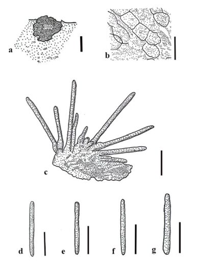
Epithamnolia is a lichenicolous genus introduced by Zhurbenko (2012), based on morphology, with the type species E. karatyginii, found on Thamnolia species in Canada. Epithamnolia is characterized by pycnidial, rugose, irregularly subglobose, blackish conidiomata becoming cupuliform with age. Conidiomata are initially closed, with an irregular opening, sub-immersed in thalli to finally superficial, arising singly and dispersed. The exciple is dark to medium brown throughout, with textura angularis, textura globulosa or textura porrecta cells. Conidiophores are absent, and conidiogenous cells line the pycnidial cavity. The conidiogenous cells are enteroblastic, phialidic, with indistinct collarette, with periclinal thickening, not proliferating, hyaline, and lageniform to narrowly elliptic and smooth-walled. Conidia are acrogenous that arise singly, not catenate, hyaline, cylindrical, with obtuse to sometimes slightly truncated ends, smooth-walled, sometimes with conspicuous guttules and septate but not constricted at the septum (Zhurbenko 2012). In the group of non-lichenicolous pycnidial genera, Epithamnolia shares similar morphological characters with Ascochyta, Clypeopycnis, Didymochaeta, Gelatinopycnis, Pocillopycnis, Pseudocenangium, and Septopatella species (Zhurbenko 2012). However, based on the following characters, these genera can be distinguished from Epithamnolia, such as having non-cupulate conidiomata (Ascochyta, Clypeopycnis, Didymochaeta), closed conidiomata with a complex wall (Gelatinopycnis), presence of the conidiophores (Clypeopycnis, Gelatinopycnis, Pocillopycnis, Pseudocenangium, Septopatella), conidiogenous cells with distinct collarette, and a periclinal thickening (Didymochaeta), sympodially proliferating conidiogenous cells (Septopatella, Pocillopycnis) and holoblastic, lunate to sigmoid, 3–10-septate conidia (Pocillopycnis), and annellidic conidiogenous cells (Pseudocenangium) (Zhurbenko 2012). Considering other coelomycetous lichenized fungi, Epithamnolia shares similar characteristics to species in Hainesia by having cupulate conidiomata and hyaline, cylindrical, septate conidia. However, Zhurbenko (2012) considered Epithamnolia and Hainesia as two different genera, distinguishing them based on the presence of branched conidiophores in Hainesia. Suija et al. (2017) mentioned that certain lichenicolous fungi belonging to Hainesia are phylogenetically not related to the genus, and proposed to include the species in Epithamnolia despite the presence of distinct conidiophores. Consequently, several Hainesia species have been transferred into Epithamnolia including H. brevicladoniae, H. longicladoniae, H. pertusariae, and H. xanthoriae (Suija et al. 2017), H. atrolazulina (Diederich et al. 2018). Additionally, two new species were introduced, E. rangiferinae (Suija et al. 2017) and E. rugosopycnidiata (Flakus et al. 2019). This genus is listed under Mniaeciaceae, Leotiales (Leotiomycetes) (Wijayawardene et al. 2022). Currently, there are eight species listed in Index Fungorum (2023), and these species are reported on different hosts from different countries (Russia, different parts of Europe, and America).
Type species: Epithamnolia karatyginii Zhurb.
Other accepted species – (Species Fungorum – search Epithamnolia)
- Epithamnolia brevicladoniae (Diederich & van den Boom) Diederich & Suija
- Epithamnolia longicladoniae (Diederich & van den Boom) Diederich & Suija
- Epithamnolia pertusariae (Etayo & Diederich) Diederich & Suija
- Epithamnolia rangiferinae E. Zimm., Diederich & Suija
- Epithamnolia xanthoriae (Brackel) Diederich & Suija
- Epithamnolia atrolazulina (Etayo) Diederich
- Epithamnolia rugosopycnidiata Etayo & Flakus
Figure 1 – Epithamnolia karatyginii (LE 260498, holotype). a Conidiomata. b Exciple in water. c Conidiogenous cells and conidia in water. d-g Conidia. Scale bars: a = 100 μm, b-g = 10 μm. Redrawn from Zhurbenko (2012).
References
Entry by
Ashani D. Madagammana, Center of Excellence in Fungal Research, Mae Fah Luang University, Chiang Rai, 57100, Thailand; School of Science, Mae Fah Luang University, Chiang Rai, 57100, Thailand.
(Edited by Chitrabhanu S. Bhunjun, Subodini N. Wijesinghe, Samaneh Chaharmiri-Dokhaharani, & Achala R. Rathnayaka)
Published online 9 January 2024
