Fungalpedia – Note 74 Elongaticonidia
Elongaticonidia W.J. Li, E. Campores & K.D. Hyde
Citation when using this data: Huanraluek et al. (in prep), Fungalpedia, coelomycetes
Index Fungorum, Facesoffungi, MycoBank, GenBank, Coleomycetes.org, Fig1.
Elongaticonidia was introduced as a saprobe on dead spines of Rosa canina from Italy (Li et al. 2020), and is a monotypic genus until today. Phylogenetic analyses based on LSU sequence data showed that Elongaticonidia is closer to Mulderomyces, but it formed a separate branch in Ostropales (Lecanoromycetes) (Li et al. 2020, Wijayawardene et al. 2021). Conidiomata are black, pycnidial, solitary to gregarious, subepidermal, immersed, globose to subglobose and ostiolate. Ostioles are short and centrally located. The conidiomata wall is composed of thick-walled, dark brown to brown cells of textura angularis. The conidiophores are reduced to conidiogenous cells. Conidiogenous cells form from the inner layer of the conidiomata, are pale brown to hyaline, holothallic, ampulliform to cylindrical, discrete, determinate, smooth-walled, consisting of basal thicker-walled cells which become flared at the inception of conidiogenesis. Conidia are hyaline, arthric, formed by disarticulation of the conidial chain, produced in simple unbranched chains with the youngest conidium at the base, elongated, 1-septate, deeply constricted at the septum, bearing unbranched, cellular flexuous, appendages. The morphological characteristics of this genus are different from Mulderomyces in the form of conidia and conidiogenous cells; Mulderomyces has proliferating sympodial, subcylindrical conidiogenous cells, and cylindrical, 2–6-septae, prominently constricted at septa, with mature conidia breaking into phragmospores (Crous et al. 2016, Li et al. 2020).
Types species: Elongaticonidia rosae W.J. Li & Camporesi & K.D. Hyde 2020
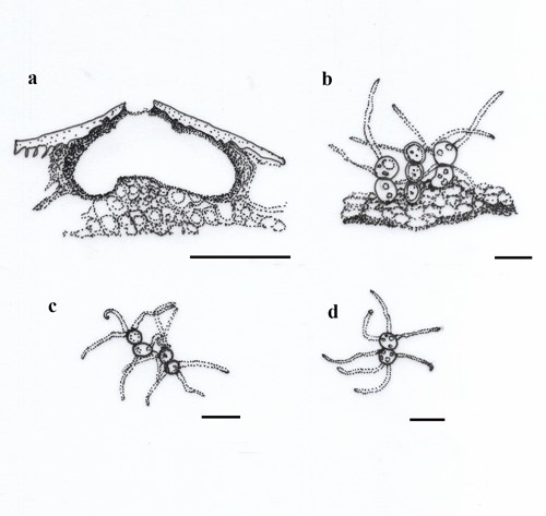
Fig 1 – Elongaticonidia rosae (redrawn from Li et al. 2020) a Vertical section of conidiomata. b Conidiogenous cells and developing conidia. c, d Conidia. Scale bars: a = 100 µm, b = 10 µm, c, d = 5 µm.
References
Crous PW, Wingfield MJ, Burgess TI, Le Roux JJ, et al. 2016 – Fungal Planet description sheets: 400–468. Persoonia 36,316–458.
https://doi.org/10.3767/003158516X692185
Li WJ, McKenZie EHC, Liu JK, Bhat DJ, et al. 2020 – Taxonomy and phylogeny of hyaline-spored coelomycetes. Fungal Diversity 100, 279–801.
https://doi.org/10.1007/s13225-020-00440-y
Wijayawardene NN, Hyde KD, Anand G, Dissanayake LS, et al. 2021 – Towards incorporating asexually reproducing fungi in the natural classification and notes for pleomorphic genera. Mycosphere 12(1), 238–405.
https://doi.org/10.5943/mycosphere/12/1/4
Entry by
Naruemon Huanraluek, Center of Excellence in Fungal Research, Mae Fah Luang University, Chiang Rai, Thailand.
(Edited by Kevin D Hyde and Ruvishika S. Jayawardena)
Published online 14 September 2023