Fungalpedia – Note 275, Distoseptispora
Distoseptispora K.D. Hyde, McKenzie & Maharachch.
Citation when using this entry: Perera et al. 2024 (in prep) – Fungalpedia, genera described in 2016.
Index Fungorum, Facesoffungi, MycoBank, GenBank, Fig. 1
Classification: Distoseptisporaceae, Distoseptisporales, Incertae sedis, Sordariomycetes, Pezizomycotina, Ascomycota, Fungi
Distoseptispora, a sporidesmium-like hyphomycetous genus, was introduced by Su et al. (2016) based on the LSU-SSU-ITS phylogeny. The genus accommodates D. fluminicola as the type species. The generic concept of Distoseptispora was emended by Yang et al. (2018). Distoseptispora is characterized by superficial, effuse, hairy, or velvety black colonies on the substratum. The mycelium is mostly immersed and composed of branched, septate, smooth-walled, and pale brown hyphae. Macronematous mononematous conidiophores occur either singly or in groups. They are unbranched, septate, erect, straight or flexuous, smooth-walled, olivaceous to brown, cylindrical, and robust at the base. Conidiogenous cells are monoblastic, integrated, determinate, terminal, cylindrical, and sometimes with percurrent proliferation. Acrogenous conidia are solitary, olivaceous, brown or yellowish/reddish brown and euseptate or distoseptate. They are obclavate or cylindrical in shape, with a rounded apex and a truncate base. The conidial length is indeterminate or exhibited percurrent proliferation. Basal cell of conidia with cross wall and basal scar. The conidial secession is schizolytic. The sexual morphs of this genus remain undetermined (Su et al. 2016; Yang et al. 2018). There are 74 species listed in Index Fungorum (2024) under Distoseptispora. Distoseptispora species are commonly found in freshwater habitats (Yang et al. 2021; Zhang et al. 2022).
Type species: Distoseptispora fluminicola McKenzie, Hong Y. Su, Z.L. Luo & K.D. Hyde
Other accepted species: Species Fungorum – search Distoseptispora
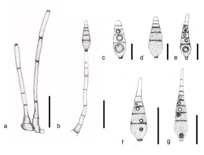
Figure 1 – Distoseptispora fluminicola (HKAS 84006, holotype). a, b Conidiophores and conidia. c–g Conidia. Scale bars: a = 30 μm, b = 70 μm, c, e, f = 50 μm, d, g = 70 μm. Redrawn from Su et al. (2016).
References
Entry by
Rekhani Hansika Perera, Center of Excellence in Fungal Research, Mae Fah Luang University, Chiang Rai, 57100, Thailand.
(Edited by Kevin D. Hyde, Samaneh Chaharmiri-Dokhaharani, & Achala R. Rathnayaka)
Published online 28 May 2024