Fungalpedia – Note 551, Dinomyces
Dinomyces Karpov & Guillou
Citation when using this data: Tibpromma et al. 2024 (in prep.) – Fungalpedia, Chytridiomycota.
Index Fungorum, Facesoffungi, MycoBank, GenBank, Fig. 1
Classification: Dinomycetaceae, Rhizophydiales, Incertae sedis, Rhizophydiomycetes, Chytridiomycotina, Chytridiomycota, Fungi.
Dinomyces (Dinomycetaceae) was established by Lepelletier et al. (2014) as a monotypic genus with the type species Dinomyces arenysensis based on phylogenetic and morphological characteristics studies. This species is the first chytrid known to infect marine dinoflagellates, instead there are many chytrids infecting microalgae in freshwater (Gleason et al. 2011). Dinomyces is characterized by a parasitoid of marine dinoflagellates with a simple thallus with inoperculate, monocentric, epibiotic sporangium with endogenous development, apophysum, and branching rhizoidal axis (Lepelletier et al. 2014). Zoospores are distinguished by a central ribosomal aggregation that separates the nucleus from the microbody-lipid complex (MLC), which comprises a solitary microbody surrounding a sizable lateral lipid globule with fenestrated cisterna. MLC are associated with a single mitochondrion. Small, dense structures were observed in the peripheral cytoplasm. The kinetid is positioned adjacent to the ribosomal core. The flagellar transition zone includes a spiral fiber. The lateral root, which consists of five microtubules, extends from the kinetosome to the fenestrated cisterna (Lepelletier et al. 2014).
Type species: Dinomyces arenysensis S.A. Karpov & L. Guillou
Other accepted species: Species Fungorum – search Dinomyces
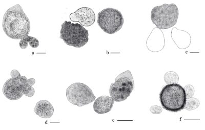
Figure 1 Infection by Dinomyces arenysensis. a Early infection in Ostreopsis cf. ovata, host cell starts to become granulated inside. b Late infection in O. cf. ovata, the cytoplasm of the host cell became black, compared with a healthy cell in the same picture. c Fungus sporangia infecting O. cf. ovata stained with calcofluor under epifluorescence microscopy. d Alexandrium andersonii, polyinfection. e Infection on one strain of Scrippsiella trochoidea vegetative cell. f Infection of S. trochoidea resting cyst (same strain as precendently). Scale bars=10 μm. Redrawn from Lepelletier et al. (2014).
References
Entry by
Lu W, Excellence Center of Microbial Diversity and Sustainable Utilization, Department of Biology, Faculty of Science, Chiang Mai University, Chiang Mai 50200, Thailand; Center for Yunnan Plateau Biological Resources Protection and Utilization, College of Biological Resource and Food Engineering, Qujing Normal University, Qujing, Yunnan 655011, China
(Edited by Saowaluck Tibpromma, Samaneh Chaharmiri-Dokhaharani, & Achala R. Rathnayaka)
Published online 13 December 2024
