Fungalpedia – Note 298, Corticimorbus
Corticimorbus S.F. Chen & M.J. Wingf
Citation when using this entry: Perera et al. 2024 (in prep) – Fungalpedia, genera described in 2016.
Index Fungorum, Facesoffungi, MycoBank, GenBank, Fig. 1
Classification: Cryphonectriaceae, Diaporthales, Diaporthomycetidae, Sordariomycetes, Pezizomycotina, Ascomycota, Fungi
Chen et al. (2016) established monotypic genus Corticimorbus with C. sinomyrti as the type. The taxon was placed in Cryphonectriaceae based on LSU, 5.8S rDNA and the exon regions of the BT2 and BT1 (Chen et al. 2016). Corticimorbus is characterized by gregarious or single, superficial to slightly immersed, orange to umber and pulvinate ascostromata on bark. Cells of the ascostromatic tissue are forming a textura globulosa and covering the tops of the perithecial bases. Perithecia are fuscous black, valsoid with globose to sub-globose bases. They are embedded beneath the surface of bark at base of stromata with long, black necks extending above the stromatic surface. Perithecial necks are emerging at stromatic surface as black ostioles with orange stromatic tissue of textura porrecta. Asci are unitunicate, 8-spored, fusoid to oval and non-stipitate. They are released from inner wall of perithecia when mature. Ascospores are biseriate, hyaline, fusoid to ellipsoidal with rounded to slightly tapered ends, 1-septate and have a constriction at septum. Septum can be variously placed in the spore, but it is usually central. Conidiomata are component of ascostromata as conidial locules or as separate structures. They are black, superficial, conical to globose, without necks, uni- to multilocular structures, with locules often convoluted, stromatic tissue textura globulosa. Conidiophores are hyaline, aseptate, cylindrical, occasionally septate and branched. Conidiogenous cells are cylindrical or flask-shaped with attenuated apices. Paraphyses are absent. Conidia are hyaline, fusoid to oval and aseptate (Chen et al. 2016).
Type species: Corticimorbus sinomyrti S.F. Chen, F.F. Liu & M.J. Wingf.
Other accepted species: This genus is monotypic.
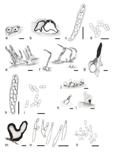
Figure 1 – Corticimorbus sinomyrti (a–d Structures from naturally occurring specimens, e–i Sexual fruiting structures induced to form on Rhodomyrtus tomentosa branch sections on MEA, k–o Asexual fruiting structures induced to form on R. tomentosa branch sections on MEA). a Ascostroma on bark. b Longitudinal section through ascostroma. c Ascus. d Ascospores. e, f Induced ascostomata on bark. g Longitudinal section through ascostroma showing stromatal tissue and neck. h Ascus. i Ascospores. j Conidiomata on the bark produced conidial spore mass. k Superficial conidioma. l Fruiting structures producing conidia in the form cirrhi. m Longitudinal section through conidioma showing black stroma. n Conidiophores and conidigenous cells. o Conidia. Scale bars: a, e, f = 500 μm, b, g = 200 μm, c, d, h, i = 10 μm, j = 500 μm, k, l = 200 μm, m = 100 μm, n, o = 10 μm. Redrawn from Chen et al. (2016).
Reference
Entry by
Rekhani Hansika Perera, Center of Excellence in Fungal Research, Mae Fah Luang University, Chiang Rai, 57100, Thailand.
(Edited by Kevin D. Hyde, Samaneh Chaharmiri-Dokhaharani, & Achala R. Rathnayaka)
Published online 8 July 2024