Fungalpedia – Note 404, Cercopemyces
Cercopemyces T.J. Baroni, Kropp & V.S. Evenson
Citation when using this data: Tibpromma et al. 2024 (in prep.) – Fungalpedia, Macrofungi.
Index Fungorum, Facesoffungi, MycoBank, GenBank, Fig. 1
Classification: Incertae sedis, Agaricales, Agaricomycetidae, Agaricomycetes, Agaricomycotina, Basidiomycota, Fungi.
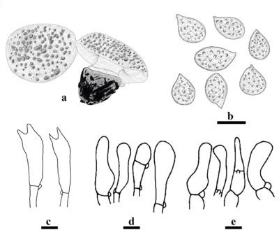
Based on morphology and phylogeny of LSU, ITS and RPB1, Baroni et al. (2014) introduced Cercopemyces to accommodate the type species Cercopemyces crocodilinus, and Cercopemyces ponderosus. Dima (2015) introduced Cercopemyces rickenii and basionymized it with Armillaria rickenii. Currently, only three species are accepted in this genus, which are found in soils in Europe and the USA (Baroni et al. 2014, Dima 2015, Sukhomlyn et al. 2019). Cercopemyces is characterized by large fleshy amanitoid robust basidiomata with thick pyramidal scales or conical warts covering the pileus, narrowly clavate basidia with long and tapered sterigmata, and ellipsoid, broadly ellipsoid to globose basidiospores with a delicately punctate roughened ornamentation (Baroni et al. 2014). Phylogenetically, Cercopemyces is related to Ripartitella and Cystodermella. However, Cercopemyces differs from Ripartitella by the latter having robust basidomata, melanoleuca-type pleurocystidium that is lageniform or ampullaceous and crystalline encrusted, basidiospores with obvious echinulate ornamentations, and the habits that occur on decaying wood or woody debris (Baroni et al. 2014). Cystodermella is differs from Cercopemyces by reddish-brown cap with conical granules and warts, and absent cystidia (Harmaja 2002, Kuo 2004). Cercopemyces can be distinguished from other genera based on morphology and multigene phylogeny.
Type species: Cercopemyces crocodilinus T.J. Baroni, Kropp & V.S. Evenson
Other accepted species: Species Fungorum – search Cercopemyces
Figure 1 – Morphological features of Cercopemyces crocodilinus. a Basidiomata. b Basidiospores. c Basidia. d Cystidiate end cells from appendiculate tissue on pileus margin. e Cystidiate end cells from annular ring on stipe basal bulb. Scale bars: b,c = 5 μm, d,e = 10 μm. Redrawn from Baroni et al. (2014).
References
Entry by
Liu XF, Center for Yunnan Plateau Biological Resources Protection and Utilization, College of Biological Resource and Food Engineering, Qujing Normal University, Qujing, Yunnan 655011, China; Center of Excellence in Fungal Research, Mae Fah Luang University, Chiang Rai 57100, Thailand; School of Science, Mae Fah Luang University, Chiang Rai 57100, Thailand.
(Edited by Saowaluck Tibpromma, Samaneh Chaharmiri-Dokhaharani, & Achala R. Rathnayaka)
Published online 26 November 2024
