Fungalpedia – Note 309, Capitofimbria
Capitofimbria L. Lombard & Crous
Citation when using this entry: Perera et al. 2024 (in prep) – Fungalpedia, genera described in 2016.
Index Fungorum, Facesoffungi, MycoBank, GenBank, Fig. 1
Classification: Stachybotryaceae, Hypocreales, Hypocreomycetidae, Sordariomycetes, Pezizomycotina, Ascomycota, Fungi
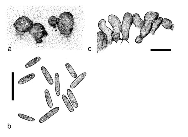
Lombard et al. (2016) established the monotypic genus Capitofimbria for Myrothecium compactum, which clustered distant to the Myrothecium s.str. clade in the analysis of cmdA, ITS, rpb2, and tub2 loci. Capitofimbria is characterized by sporodochial conidiomata that occur superficially on the substrate, and are stromatic, scattered, or rarely gregarious. Conidiomata are oval to irregular in outline, amphigenous, pulvinate, with olivaceous green to dark green slimy mass of conidia and lack a white setose fringe surrounding the conidial mass. Stromas are hyaline to subhyaline and well-developed with globulosa and angularis texture. Marginal hyphae that surround the sporodochia are septate, branched, or unbranched, and terminated in a thick-walled, capitate-to-clavate cell. The marginal hyphae are compactly crowded, coarsely rugose or tuberculate, pale brown-green and that turns dark brown-green at the apex. The conidiophores are macronematous, tightly aggregated, septate with smooth walls, and have a subhyaline to pale olivaceous brown apex. Conidiogenous cells are phialidic, cylindrical to slightly subulate, aseptate and smooth-walled. They had prominent collarette and periclinal thickening. Conidia are aseptate, olivaceous brown, cylindrical, rounded at both ends with smooth walls. The sexual morph remains undetermined (Castañeda-Ruíz et al. 2008; Lombard et al. 2016). Capitofimbria compacta is associated with dead plant parts (Castañeda-Ruíz et al. 2008; Lombard et al. 2016).
Type species: Capitofimbria compacta (R.F. Castañeda, Gusmão, Stchigel & M. Stadler) L. Lombard & Crous
Other accepted species: This genus is monotypic.
Figure 1 – Capitofimbria compacta (CBS 111739, ex-type). a Sporodochial conidiomata on SNA. b Conidia. c Marginal hyphae of the sporodochia. Scale bars: b, c = 10 μm. Redrawn from Lombard et al. (2016).
References
Entry by
Rekhani Hansika Perera, Center of Excellence in Fungal Research, Mae Fah Luang University, Chiang Rai, 57100, Thailand.
(Edited by Kevin D. Hyde, Samaneh Chaharmiri-Dokhaharani, & Achala R. Rathnayaka)
Published online 27 August 2024
