Fungalpedia – Note 212, Ascochalara
Ascochalara Réblová.
Citation when using this entry: Silva et al. 2024 (in prep) – Fungalpedia, Sordariomycetidae.
Index Fungorum, Facesoffungi, MycoBank, GenBank, Fig. 1
Classification: Chaetosphaeriaceae, Chaetosphaeriales, Sordariomycetidae, Sordariomycetes, Pezizomycotina, Ascomycota, Fungi
Ascochalara was introduced by Réblová (1999) as a monospecific genus, based on morphological characterization (Réblová 1999). Based on the perithecial wall, asci, paraphyses, and ascospores, and the chalara-like asexual morph, unique combinations of characteristics differentiate it from other known genera in Chaetosphaeriaceae (Réblová 1999). The type species, Ascochalara gabretae Réblová, was found as a saprobe underneath the bark of Abies alba, which is barely decomposed (Réblová 1999).
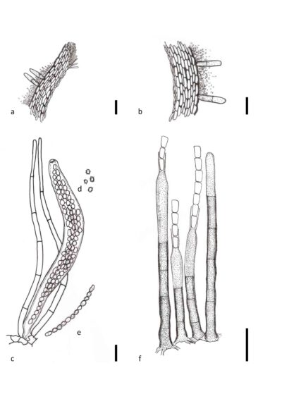
Conidiophores are dematiaceous hyphomycetes, macronematous, manonematous, unbranched, and erect, with phialide terminals that are paler than the base. The collarette is subhyaline to hyaline and cylindrical. Conidia are subcylindrical to wedge-shaped, forming short or long chains that adhering together, with a round apex with truncate base, smooth, and hyaline (Réblová 1999, Hyde et al. 2020). In contrast, the sexual morph is a superficial perithecium, black, subglobose to globose, papillate, fragile ascomata wall, and a periphysate ostiolar canal. Paraphyses are abundant and septate. Asci are cylindrical to clavate, short-stipitate, with a J- apical annulus. When young, ascospores are long-fusiform to cylindrical, gradually separating into part-spores with maturity inside the ascus, 2-3-seriate. Part-spores are nearly rounded, hyaline, smooth, or slightly verrucose (Réblová 1999, Hyde et al. 2020). Although Ascochalara was introduced in 1999, it remains a monospecific genus without any addition (Species Fungorum 2023).
Type species: Ascochalara gabretae Réblová
Figure 1 – Ascochalara gabretae. a Cross-section of ostiole. b Cross-section of perithecium. c Asci with fragmented spores and paraphyses. d Part-spores, e Ascospores after separating into part-spores. f Conidiophores and conidia adhering in chains. Scale bars: a, b = 20 µm and c–f = 10 µm. Redrawn from: Réblová (1999).
References
Entry by
Veenavee Sulakkana H. Silva, Center of Excellence in Fungal Research, Mae Fah Luang University, Chiang Rai 57100, Thailand.
(Edited by Antonio Roberto Gomes de Farias, Kevin D. Hyde, Samaneh Chaharmiri-Dokhaharani, & Achala R. Rathnayaka)
Published online 15 February 2024
