Fungalpedia – Note 355, Areospora
Areospora G.D. Stentiford, S. Bateman, S.W. Feist, S. Oyarzún, J.C. Uribe, M. Palacios & D.M. Stone
Citation when using this data: Tibpromma et al. 2024 (in prep.) – Fungalpedia, Parasites.
Index Fungorum, Facesoffungi, MycoBank, GenBank, Fig. 1
Classification: Areosporiidae, Incertae sedis, Incertae sedis, Microsporea, Incertae sedis, Microsporidia, Protozoa.
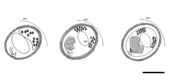
Areospora was established with Areospora rohanae as the type species. Furthermore, the phylogenetic and morphological distinction from other diverse parasite phylum, therefore established a new genus under new family Areosporiidae to accommodate this parasite species (Stentiford et al. 2014). Merogonal and sporoogonal stages, in accordance with the generic description for Areospora, are found within the phagocytes and connective tissue cells of lithodid crabs. Parasite life stages can be enclosed within a specialized parasitophorous vacuole (SPV). The early sporogony is characterized by a distinctive rosette-like structure that is confined within the SPV. Rosettes comprise 8 pre-sporonts. The transition from rosette-like sporonts to early sporoblasts is characterized by the formation of spore-extrusion precursors and the migration of tubular bristles to the surface of the pre-sporoblast exospore layer, monokaryotic spores, adorned with unique bristles (Stentiford et al. 2014).
Type species: Areospora rohanae G.D. Stentiford, S. Bateman, S.W. Feist, S. Oyarzún, J.C. Uribe, M. Palacios & D.M. Stone
Other accepted species: Species Fungorum – search Areospora
Figure 1 – Morphology of Areospora rohanae. Early to mature sporoblast with thickened endospore and ornamented spore wall (SVP=sporophorous vesicle). Scale bar = 1 μm. Redrawn from Stentiford et al. (2014).
Reference
Entry by
Yang EF, Department of Biology, Faculty of Science, Chiang Mai University, Chiang Mai 50200, Thailand; Center for Yunnan Plateau Biological Resources Protection and Utilization, College of Biological Resource and Food Engineering, Qujing Normal University, Qujing, Yunnan 655011, China.
(Edited by Saowaluck Tibpromma, Samaneh Chaharmiri-Dokhaharani & Achala R. Rathnayaka)
Published online 14 November 2024
