Fungalpedia – Note 168,Anaseptoidium
Anaseptoidium R.F. Castañeda, Heredia & R.M. Arias
Citation when using this entry: Madagammana et al. 2024 (in prep) – Fungalpedia, genera described in 2012.
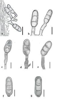
Index Fungorum, Facesoffungi, MycoBank, GenBank, Fig 1
Classification: Pezizomycotina, Ascomycota, Fungi.
Anaseptoidium, an asexual genus introduced by Castañeda-Ruiz et al. (2012), based on morphology, accommodates A. mycophilum as the type species. Due to the lack of sequence data, this genus is listed under the Ascomycota genera incertae sedis (Wijayawardene et al. 2022). Hence, additional taxon sampling and sequence data are necessary to confirm the phylogenetic position of this genus. Anaseptoidium is characterized by effuse, creeping, and brown colonies on the natural substrate. Mycelia are superficial and immersed, with branched and septate hyphae, while stomatopodia are absent. Conidiophores are macronematous, mononematous, erect, and pale brown, sometimes reduced to monoblastic, integrated, and determinate conidiogenous cells (Castañeda-Ruiz et al. 2021). Schizolytic conidial secession produces solitary, acrogenous, cylindrical to oblong, fimbriate, septate, smooth, or verruculose conidia, which are pale brown to brown. Anaseptoidium is similar to Septoidium. However, it differs from Septoidium by having monoblastic, annellate conidiogenous cells with percurrent proliferation. Additionally, Anaseptoidium produces stomatopodia through assimilative hyphae on the substrate, distinguishing it from Septoidium. Furthermore, the secession of each conidium in Anaseptoidium is schizolytic, but unlike most closely related fungal genera, the outer and inner layers of the wall do not separate simultaneously. Currently, there is only one species listed in Index Fungorum (2023) under Anaseptoidium, and it was collected on the synnemata of Phaeoisaria clavulata in Mexico (Castañeda-Ruiz et al. 2012).
Type species: Anaseptoidium mycophilum R.F. Castañeda, Heredia & R.M. Arias.
Figure 1 – Anaseptoidium mycophilum (XAL CB1696, ex-holotype). a, b Conidiophores, conidiogenous cells, and conidia. c-e Conidiogenous cells and conidia. f, g Conidia. Scale bars: a, b = 10 µm, c-g = 5 µm. Redrawn from Castañeda-Ruiz et al. (2012)
References
Entry by
Ashani D. Madagammana, Center of Excellence in Fungal Research, Mae Fah Luang University, Chiang Rai, 57100, Thailand; School of Science, Mae Fah Luang University, Chiang Rai, 57100, Thailand.
(Edited by Chitrabhanu S. Bhunjun, Kevin D. Hyde, Subodini N. Wijesinghe, Samaneh Chaharmiri-Dokhaharani & Achala R. Rathnayaka)
Published online 9 January 2024
