Fungalpedia – Note 194, Dictyosporites (Fossil Fungi)
Dictyosporites Félix emend. Kalgutkar & Janson.
Citation when using this data: Saxena RK & Hyde KD. 2024 (in prep) – Fungalpedia, Fossil Fungi.
Index Fungorum, Facesoffungi, MycoBank, GenBank, Fig. 1
Classification: Dictyosporae, Fossil Fungi.
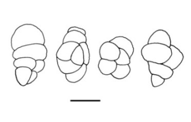
The monotypic fossil genus, Dictyosporites was instituted by Félix (1894) from the Eocene sediments of Perekeschkul, near Baku, Azerbaijan, and provided the following diagnosis: “The so-called wall-shaped conidia become multicellular by repeated transverse and longitudinal divisions. In addition to large conidia, whose growth can probably be regarded as complete, uni- and bicellular conidia representing the initial developmental stages also occur. They are all of the brownish colouration. Their outlines are rather variable, depending on the conidium’s position to the section’s plane. Viewed from the top or bottom, they often appear spherical with flatly indented outlines; longitudinal sections are irregular; elliptical, pear-shaped, or resembling short, corpulent snails (e.g., Turbo).
The maximum length is 0.0204 mm (20.4 μm), and the maximum diameter is 0.0153 mm (15.3 μm); the respective dimensions of only bicellular conidium are 0.0102 and 0.0085 mm (10.2 and 8.5 μm).” Kalgutkar & Jansonius (2000) emended the diagnosis as follows: “Inaperturate, multicellate (apparently by internal septation, of irregular pattern), muriform fungal spores, cells rounded to rounded polygonal. The overall shape is rounded, oval/ovoid to elongate; indentations may occur where septa intersect the amb. A hilum cannot be discerned. Staphlosporonites differs in showing a distinct hilum, or proximal hilar cell.” Papulosporonites consist of spore clusters or aggregates, in which there is no suggestion of linear or planar symmetry. There are 22 records included in Index Fungorum (2023) under this genus.
Synonyms: Arbusculites Paradkar, Dactylosporites Paradkar, Pleosporonites R.T. Lange & P.H. Sm., Ravenelites Ramanujam & Ramachar.
Type species: Dictyosporites loculatus Félix.
Figure 1 – Dictyosporites loculatus Félix 1894. Scale bar = 5 μm (redrawn from Félix 1894)
References
Lange RT, Smith PH. 1971 – The Maslin Bay flora, South Australia. 3. Dispersed fungal spores. Neves Jahrb. Geol. Palaontol. Monatsh. 11, 663–681.
Entry by
Ramesh K. Saxena, Birbal Sahni Institute of Palaeosciences, Lucknow, India
(Edited by Kevin D. Hyde, Samaneh Chaharmiri-Dokhahari, & Achala R. Rathnayaka)
Published online 1 February 2024
