Fungalpedia – Note 407, Pseudospiromastix
Pseudospiromastix Guarro, Stchigel & Cano
Citation when using this data: Tibpromma et al. 2024 (in prep.) – Fungalpedia, Ascomycota.
Index Fungorum, Facesoffungi, MycoBank, GenBank, Fig. 1
Classification: Incertae sedis, Incertae sedis, Eurotiomycetidae, Eurotiomycetes, Pezizomycotina, Ascomycota, Fungi.
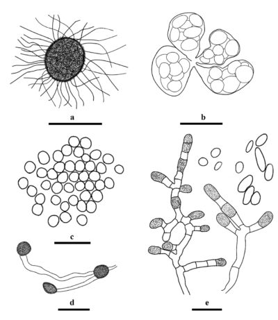
Pseudospiromastix was introduced by Rizzo et al. (2014) as a monotypic genus and typed as P. tentaculata based on the morphology and phylogeny of LSU, which is etymologically based on the genus name Spiromastix. However, the name Pseudospiromastix and the new combination P. tentaculata are nomenclaturally invalid because there is no basionym fully cited (Hirooka et al. 2016). Hirooka et al. (2016) validated Pseudospiromastix and placed it in Spiromastigaceae, based on the phylogenetic tree of SSU + LSU. Currently, only one species has been introduced in this genus, which has been isolated from soil in Europe (Italy) and Africa (Nigeria, Rwanda, and South Africa) (Rizzo et al. 2014, Hirooka et al. 2016, GBIF 2024). Pseudospiromastix is characterized by discrete, orange-brown to mid brown, globose to subglobose ascomata, subglobose or ovoid to ellipsoidal, evanescent, eight-spored asci, and pale brown, lenticular, covered with several irregularly pitted furrows ascospores, sparingly branched or laterally branched, thick-walled conidiogenous hyphae, terminal and single lateral conidia unicellular, septate, cylindrical, cuboid or doliiform, thick-walled arthroconidia, and terminal or intercalary chlamydospores (Rizzo et al. 2014, Hirooka et al. 2016), and the most distinctive sexual characteristic is the undulate ascomata appendage with swollen ends (Hirooka et al. 2016). Pseudospiromastix can be distinguished from other genera based on its morphology and phylogeny.
Type species: Pseudospiromastix tentaculata (Guarro, Gené & De Vroey) Guarro, Stchigel & Cano
Other accepted species: This genus is monotypic.
Figure 1 – Morphological features of Pseudospiromastix tentaculata. a Ascoma. b Asci. c Ascospores. d Chlamydospores. e Conidiophores and conidia. Scale bars: a-d = 10 μm. Redrawn from Hirooka et al. (2016).
References
Entry by
Liu XF, Center for Yunnan Plateau Biological Resources Protection and Utilization, College of Biological Resource and Food Engineering, Qujing Normal University, Qujing, Yunnan 655011, China; Center of Excellence in Fungal Research, Mae Fah Luang University, Chiang Rai 57100, Thailand; School of Science, Mae Fah Luang University, Chiang Rai 57100, Thailand.
(Edited by Saowaluck Tibpromma, Samaneh Chaharmiri-Dokhaharani, & Achala R. Rathnayaka)
Published online 26 November 2024
