Fungalpedia – Note 322, Sirastachys
Sirastachys L. Lombard & Crous
Citation when using this entry: Perera et al. 2024 (in prep) – Fungalpedia, genera described in 2016.
Index Fungorum, Facesoffungi, MycoBank, GenBank, Fig. 1
Classification: Stachybotryaceae, Hypocreales, Hypocreomycetidae, Sordariomycetes, Pezizomycotina, Ascomycota, Fungi.
Lombard et al. (2016) established Sirastachys for a group of stachybotrys-like taxa that form synnemata in culture with conidiophores that emerge laterally. In phylogenetic analysis based on cmdA, ITS, rpb2, and tub2 loci, Sirastachys formed a robust clade away from Stachybotrys s. str. clades (Lombard et al. 2016). Sirastachys is characterized by cylindrical synnemata that are hyaline, slender to robust and straight to curved. The synnemata are composed of bundles of parallel, longitudinal, and tightly packed hyphae. Conidiophores emerge laterally from synnemata, are macronematous mononematous erect and occur solitary or in groups. They are thin or thick, smooth to verrucose walls, unbranched or branched, 1–3-septate, and hyaline to pale olivaceous. Each conidiophore produces an apical cluster of 6–12 conidiogenous cells. Conidiogenous cells are phialidic, elongate doliiform to clavate to subclavate, smooth to slightly verrucose, hyaline to pale olivaceous, with prominent collarettes and periclinal thickenings. Species of this genus produce ellipsoidal to obovoid to cylindrical conidia with rounded ends and smooth to verrucose walls. Conidi is aseptate and hyaline to pale olivaceous brown to dark brown in color. The sexual morph remains undetermined (Lombard et al. 2016; Tibpromma et al. 2018). Sirastachys species occur on the decaying plant leaves (Lombard et al. 2016). This genus includes nine species (Index Fungorum 2024).
Type species: Sirastachys phaeospora L. Lombard & Crous
Other accepted species: Species Fungorum, search Sirastachys
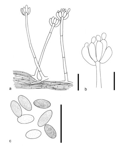
Figure 1 – Sirastachys phaeospora (CBS 100155, ex-type). a Conidiophores. b Conidiogenous cells. c Conidia. Scale bars: a = 20 μm, b, c = 10 μm. Redrawn from Lombard et al. (2016).
References
Entry by
Rekhani Hansika Perera, Center of Excellence in Fungal Research, Mae Fah Luang University, Chiang Rai, 57100, Thailand.
(Edited by Kevin D. Hyde, Samaneh Chaharmiri-Dokhaharani, & Achala R. Rathnayaka)
Published online 27 August 2024